

Review
doi: 10.1167/iovs.15-19043.
Optical Coherence Tomography Angiography
- PMID: 27409483
- PMCID: PMC4968919
- DOI: 10.1167/iovs.15-19043
Item in Clipboard
Review
Optical Coherence Tomography Angiography
Invest Ophthalmol Vis Sci.
.
Abstract
Optical coherence tomography angiography (OCTA) is a noninvasive approach that can visualize blood vessels down to the capillary level. With the advent of high-speed OCT and efficient algorithms, practical OCTA of ocular circulation is now available to ophthalmologists. Clinical investigations that used OCTA have increased exponentially in the past few years. This review will cover the history of OCTA and survey its most important clinical applications. The salient problems in the interpretation and analysis of OCTA are described, and recent advances are highlighted.
Figures

(A) The first demonstration of blood flow imaging in the living human eye with OCT. B-scan image of central vessels superior to the optic nerve head with Doppler shift signal (false color) overlaid on structural OCT (gray scale). Reprinted with permission from Yazdanfar S, Rollins AM, Izatt JA. Imaging and velocimetry of the human retinal circulation with color Doppler optical coherence tomography. Opt Lett. 2000;25:1448–1450. © 2000 Optical Society of America. (B) Amplitude-decorrelation angiography of the macula (3 × 3–mm area) using full-spectrum (left) and split-spectrum processing (right). En face angiograms represent the maximum flow projection in the inner retina slab. Split-spectrum processing reduced noise and improved visualization of the retinal vascular network.

Segmentation and processing of OCTA. Optical coherence tomography angiography was performed on a 70-kHz SD-OCT with a center wavelength of 840 nm and axial resolution of 5 μm in tissue. (A) The volumetric OCTA scan comprised 304 cross-sectional frames along the slow scan axis. Flow in each frame was computed using the SSADA algorithm. The cross-sectional angiogram shows blood flow (color) overlaid on structural OCT (gray scale). It shows that flow in inner retinal vessels (purple) are projected onto the RPE complex (indicated by white arrows) – this is called “flow projection artifact.” Image processing software separates the vitreous, inner retinal layers, outer retinal layer, and choroidal layers along the ILM and outer boundaries of the inner plexiform layer (IPL), OPL, and BM (dotted green lines). Maximum flow projection or mean reflectance projection was used to produce en face images. (B) The vitreous angiogram shows the absence of vascular flow. (C) The superficial inner retinal angiogram shows normal retinal circulation with a foveal avascular zone. Residual motion artifact in the form of a horizontal line is seen near the top of the angiogram. (D) The deep inner retina angiogram shows the deep retinal plexus. (E) The outer retina slab shows choroidal neovascularization (CNV) along with flow projection artifacts cast by the retinal circulation. (F) The choriocapillaris angiogram. (G) The deeper choroid en face structural OCT. (H) Detection of the CNV using a saliency-based approach. (I) Composite en face angiogram of the inner retina (purple) and CNV (yellow).

A case of central serous chorioretinopathy (CSCR). (A) Early-phase FA showing leakage. (B) Late-phase FA showing staining. However, FA could not determine whether the leakage was due to CSCR or secondary CNV. (C) A 3 × 3–mm en face spectral OCTA of the boxed region in panel A. (D) Cross-sectional OCTA corresponding to the green line in panel C. Optical coherence tomography angiography revealed flow beneath the RPE consistent with type 1 CNV. Optical coherence tomography angiography was useful in making the decision to treat this patient with anti-VEGF.
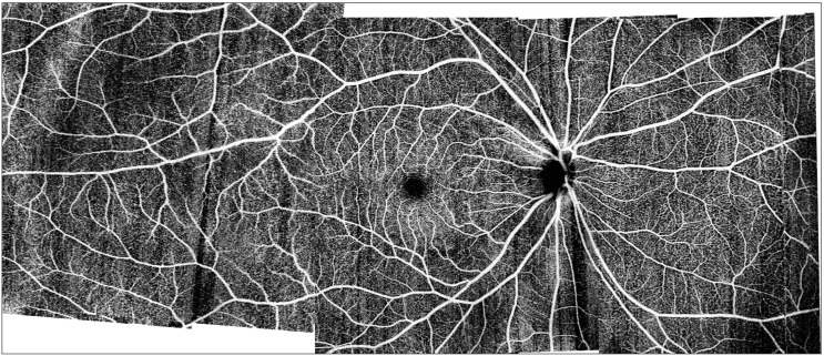
Ultra-widefield OCTA (∼20-mm width, 10-mm height, 7-mm depth) of the retinal circulation generated by montaging four scans from a 200-kHz swept-source OCT system.

(Top) Projection-resolved OCTA115 of a healthy eye shows three distinct plexuses: (A) superficial, in the nerve fiber and ganglion cell layers; (B) intermediate, between the inner plexiform and inner nuclear layers; and (C) deep, between the inner nuclear and outer plexiform layers. The plexuses merge at the edge of the foveal avascular zone. (Bottom) Optical coherence tomography angiography of an eye with nonproliferative diabetic retinopathy (NPDR) showing incongruent areas of capillary nonperfusion are present in the three plexuses. Dilated shunt vessels are seen in the intermediate (E) and deep (F) plexuses, in contrast with the uniform capillary network in the healthy eye. Reprinted with permission from Zhang M, Hwang TS, Campbell JP, et al. Projection-resolved optical coherence tomographic angiography. Biomed Opt Express 2016;7:816–828. © 2016 Optical Society of America.
References
-
- Drexler W,, Liu M,, Kumar A,, Kamali T,, Unterhuber A,, Leitgeb RA. Optical coherence tomography today: speed, contrast, and multimodality. J Biomed Opt. 2014; 19: 071412. - PubMed
-
- Hope-Ross M,, Yannuzzi LA,, Gragoudas ES,, et al. Adverse reactions due to indocyanine green. Ophthalmology. 1994; 101: 529–533. - PubMed
-
- Lopez-Saez MP,, Ordoqui E,, Tornero P,, et al. Fluorescein-induced allergic reaction. Ann Allergy Asthma Immunol. 1998; 81: 428–430. - PubMed
Publication types
MeSH terms
Grants and funding
LinkOut - more resources
Full Text Sources
Other Literature Sources
Miscellaneous
