. 2016 Aug 2;7(31):50548-50563.
doi: 10.18632/oncotarget.10500.

Stress-induced phosphoprotein-1 maintains the stability of JAK2 in cancer cells

Affiliations

Stress-induced phosphoprotein-1 maintains the stability of JAK2 in cancer cells

Chia-Lung Tsai et al. Oncotarget. .

Abstract

Overexpression of stress-induced phosphoprotein 1 (STIP1) - a co-chaperone of heat shock protein (HSP) 70/HSP90 - and activation of the JAK2-STAT3 pathway occur in several tumors. Combined treatment with a HSP90 inhibitor and a JAK2 inhibitor exert synergistic anti-cancer effects. Here, we show that STIP1 stabilizes JAK2 protein in ovarian and endometrial cancer cells. Knock-down of endogenous STIP1 decreased JAK2 and phospho-STAT3 protein levels. The N-terminal fragment of STIP1 interacts with the N-terminus of JAK2, whereas the C-terminal DP2 domain of STIP1 mediates the interaction with HSP90 and STAT3. A peptide fragment in the DP2 domain of STIP1 (peptide 520) disrupted the interaction between STIP1 and HSP90 and induced cell death through JAK2 suppression. In an animal model, treatment with peptide 520 inhibited tumor growth. In summary, STIP1 modulates the function of the HSP90-JAK2-STAT3 complex. Peptide 520 may have therapeutic potential in the treatment of JAK2-overexpressing tumors.

Keywords: JAK2; STIP1; cancer.


Conflict of interest statement

The authors declare no conflicts of interest.

Figures

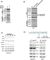
Figure 1
Figure 1. STIP1 maintains JAK2 protein stability in cancer cells
A. Ovarian (MDAH2774 and SKOV3) and endometrial (ARK2) cancer cells were transfected with control or STIP1 siRNA. JAK2 and phospho-STAT3 protein levels were determined by western blot. Actin levels were used to normalize the input proteins. B. MDAH2774 cells were transfected using the mentioned STATs (GAS1/STAT1, STAT3, and STAT5) reporter constructs, transfected with control or STIP1 siRNA, and treated with IL-6 (50 ng/mL) for 24 h. The reporter activity was measured using the luciferase assay. Results are means ± standard errors from three independent experiments. Statistical significance was calculated with the Student's t-test, *P <0.05. C. MDAH2774 and ARK2 cells were transfected with control or STIP1 siRNA. STIP1 and JAK2 RNA levels were measured with real-time quantitative PCR at 72 h after siRNA transfection. Results are means ± standard errors from three independent experiments. GAPDH was used for normalization. D. Cancer cells (MDAH2774, ARK2, and SKOV3) were treated with MG132 (25 μM) for 6 h either in the presence or absence of STIP1 siRNA. JAK2 and STIP1 protein levels were determined by western blot. E. Cancer cells (ARK2 and MDAH2774) either with or without STIP1 knockdown were transfected with His-tagged ubiquitin (His-Ub). His-Ub-labeled were immunoprecipitated with nickel beads. JAK2 levels were determined by western blot. The same quantity of protein lysates (50 μg) was used as loading control and probed with anti-JAK2, anti-STIP1, and anti-actin antibodies.
Figure 2
Figure 2. STIP1 is essential for the formation of the JAK2-HSP90-STAT3 complex
A. Endogenous STIP1 protein complexes obtained from protein lysates (2 mg) of ovarian cancer cells (MDAH2774 and SKOV3) were immunoprecipitated with anti-STIP1 or control IgG antibodies. The protein associations of halo-STIP1 with HSP90, JAK2, or STAT3 were determined with western blot. B. MDAH2774 cells were grown on coverslips. Cells were permeabilized, and an in situ proximity ligation assay (PLA) was performed to investigate protein interactions (red dots). To this aim, anti-STIP1 and anti-HSP90 (left-upper panel), anti-STIP1 and anti-STAT3 (middle-upper panel), and anti-STIP1 and anti-JAK2 (right-upper panel) antibodies were used. An IgG was used as a negative control in place of the anti-STIP1 antibody (lower panel). C, D. The truncated STIP1 constructs used for the study are shown in left panels. They included the C-terminal truncated halo-tagged STIP1s (FL: full length, R3: DP2 deleted, R2: TPR2B-DP2 deleted, and R1: DP1-TPR2A-TPR2B-DP2 deleted) (C) and the N-terminal truncated halo-tagged STIP1s (FL: full length, F3: TPR1 deleted, F2: TPR1-DP1-TPR2A deleted and F1: TPR1-DP1-TPR2A-TPR2B deleted) (D). ARK2 and 293 cells were co-transfected with the reported truncated constructs of STIP1, Flag-JAK2, and EGFP-STAT3, and subsequently purified with Halo-tag resin. Co-immunoprecipitated HSP90, JAK2, and STAT3 were analyzed with western blot using anti-HSP90, anti-Flag, and anti-EGFP antibodies, respectively. E. ARK2 and 293 cells were co-transfected with full-length NTAP-JAK2 or its truncated constructs (JAK2-840, JAK2-521) and EGFP-STAT3. Co-immunoprecipitated HSP90, STIP1, and STAT3 were analyzed with western blot using specific antibodies. NTAP-JAK2 constructs were detected using an anti-calmodulin binding peptide (CBP) antibody. NS denotes non-specific band detected with the CBP antibody. FL: full-length JAK2, 840: protein of the JAK2-840 construct, 251: protein of the JAK2-251 construct.
Figure 3
Figure 3. The interaction between STIP1 and HSP90 plays an important role in JAK2 stability
A. MDAH2774 cells were co-transfected with EGFP-STAT3 and Flag-JAK2 expression vectors; STIP1 expression was subsequently knocked down with STIP1 siRNA. Resultant cells were treated with MG132 and immunoprecipitated with anti-HSP90. The proteins involved in the formation of HSP90 complexes (STIP1, JAK2, and STAT3) were identified with western blot using specific antibodies. B. 293 cells were co-transfected with NTAP-HSP90 or the corresponding form bearing a deletion of five amino acids (720−724) in the C-terminus (NTAP-ΔMEEVD/HSP90), Flag-JAK2, and EGFP-STAT3. Pulled-down proteins were analyzed with specific antibodies. C. MDAH2774 cells were treated for 24 h with different concentrations of Novobiocin, a HSP90 C-terminal inhibitor. Protein levels of JAK2, HSP90, STIP1, and phospho-STAT3 were determined with western blot. D. The upper panel shows the positions of Antp-TPR peptide, peptide 445, and peptide 520 in STIP1. MDAH2774 cells were transiently transfected with peptide 445 or peptide 520 for 24 or 48 h. They were subsequently analyzed with western blot using the reported antibodies.
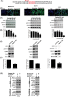
Figure 4
Figure 4. Peptide 520 blocks the JAK2-STAT3-Bcl XL pathway and induces cell death in cancer cells
A. Peptide 520 was synthesized with eight D-arginine followed by the myc-tag sequence in the N-terminus. The scramble peptide was synthesized using the same 24 amino acids of the peptide 520 in random order. B. MDAH2774 (left panel), SKOV3 (middle panel), and ARK2 (right panel) cells were transfected with peptide 520 and the sramble peptide. They were subsequently analyzed with immunofluorescence microscopy using an anti-myc antibody. C, D. MDAH2774 (left panel), SKOV3 (middle panel), and ARK2 (right panel) cells were treated with different concentrations of peptide 520. JAK2 and phospho-STAT3 protein levels were determined with western blot (C). Cell survival was investigated with the MTT assay (D). E, F. MDAH2774 (left panel), SKOV3 (middle panel), and ARK2 (right panel) cells were transfected with either peptide 520 or scramble peptide (20 μM). Protein levels of JAK2, phospho-STAT3, and Bcl XL were determined with western blot (E). Cell survival was investigated with the MTT assay (F). G. Compared with exposure to the scramble peptide (10 μM) for 24 h, peptide 520 interfered with the formation of the JAK2-STIP1-HSP90-STAT3 complex.
Figure 5
Figure 5. STIP1 and JAK2 are co-expressed in ovarian cancer tissues
A. STIP1 and JAK2 protein levels in representative ovarian cancer tissues were determined by immunohistochemistry. Tumors with high and low STIP1 expression are shown in the upper and lower panels, respectively. The scale bar represents 100 μm. B. STIP1 and JAK2 immunostaining was analyzed with a histoscore. The correlation between STIP1 and JAK2 histoscores in ovarian serous carcinomas (n = 24) is reported. C. A proximity ligation assay (PLA) was performed to investigate protein interactions in representative ovarian cancer tissues. To this aim, anti-STIP1 and anti-STAT3 (left-upper panel), anti-STIP1 and anti-JAK2 (middle-upper panel), and anti-STIP1 and anti-HSP90 (right-upper panel) antibodies were used. An IgG was used as a negative control in place of the first antibody (lower panel).
Figure 6
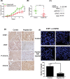
Figure 6. Peptide 520 blocks the JAK2-STAT3 pathway and inhibits tumor growth in mice
A, B. MDAH2774 cells (1 × 10⁶) were subcutaneously injected into nude mice. When a tumor volume of 10 mm3 was reached, mice were injected with 50 μL PBS (vehicle alone) or 25 μg/50 μL peptide 520 directly into the tumor (three times per week). Tumor growth was measured on a weekly basis after treatment was started. Data on volume (A) and weight (B) are expressed as means ± standard. *P <0.05 and ** P <0.01, Student's t-test. C, D. Whole cell lysates from individual tumors were obtained from experimental mice. The JAK2, phospho-STAT3, and total STAT3 protein levels were determined with western blot (C) and immunohistochemistry (D). The scale bar represents 20 μm. E. The interactions between STIP1 and HSP90 were analyzed in representative tumors in nude mice (control group and peptide 520-treated group) with the proximity ligation assay. The left panel displays a higher magnification of the right panel. The signals were counted with Image J (https://imagej.nih.gov/ij/) (lower panel).
Figure 7
Figure 7. Model showing how the HSP90-STIP1 complex controls JAK2 kinase stability
A. Summary of STIP1 domains that interact with JAK2, HSP90, and STAT3. B. STIP1 directly interacts with JAK2 and favors its delivery to HSP90 for proper protein folding, ultimately inhibiting its ubiquitin-dependent degradation in the proteasome (upper panel). Peptide 520 of the STIP1 DP2 domain blocks the interaction between STIP1 and HSP90, ultimately promoting JAK2 degradation (lower panel). Shadows of HSP90 indicated that molar ratios of these proteins were not determined in this study.

Similar articles

Cited by

References

    1. Odunuga OO, Longshaw VM, Blatch GL. Hop: more than an Hsp70/Hsp90 adaptor protein. Bioessays. 2004;26:1058–1068. - PubMed
    1. Nicolet CM, Craig EA. Isolation and characterization of STI1, a stress-inducible gene from Saccharomyces cerevisiae. Mol Cell Biol. 1989;9:3638–3646. - PMC - PubMed
    1. Carrigan PE, Riggs DL, Chinkers M, Smith DF. Functional comparison of human and Drosophila Hop reveals novel role in steroid receptor maturation. J Biol Chem. 2005;280:8906–8911. - PubMed
    1. Scheufler C, Brinker A, Bourenkov G, Pegoraro S, Moroder L, Bartunik H, Hartl FU, Moarefi I. Structure of TPR domain-peptide complexes: critical elements in the assembly of the Hsp70-Hsp90 multichaperone machine. Cell. 2000;101:199–210. - PubMed
    1. Schmid AB, Lagleder S, Grawert MA, Rohl A, Hagn F, Wandinger SK, Cox MB, Demmer O, Richter K, Groll M, Kessler H, Buchner J. The architecture of functional modules in the Hsp90 co-chaperone Sti1/Hop. EMBO J. 2012;31:1506–1517. - PMC - PubMed

MeSH terms