

The dual role of asporin in breast cancer progression
- PMID: 27409832
- PMCID: PMC5239534
- DOI: 10.18632/oncotarget.10471
The dual role of asporin in breast cancer progression
Abstract
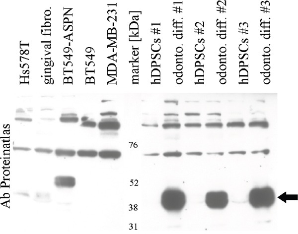
Asporin has been reported as a tumor suppressor in breast cancer, while asporin-activated invasion has been described in gastric cancer. According to our in silico search, high asporin expresion associates with significantly better relapse free survival (RFS) in patients with low-grade tumors but RFS is significantly worse in patients with grade 3 tumors. In line with other studies, we have confirmed asporin expression by RNA scope in situ hybridization in cancer associated fibroblasts. We have also found asporin expression in the Hs578T breast cancer cell line which we confirmed by quantitative RT-PCR and western blotting. From multiple testing, we found that asporin can be downregulated by bone morphogenetic protein 4 while upregulation may be facilited by serum-free cultivation or by three dimensional growth in stiff Alvetex scaffold. Downregulation by shRNA inhibited invasion of Hs578T as well as of CAFs and T47D cells. Invasion of asporin-negative MDA-MB-231 and BT549 breast cancer cells through collagen type I was enhanced by recombinant asporin. Besides other investigations, large scale analysis of aspartic acid repeat polymorphism will be needed for clarification of the asporin dual role in progression of breast cancer.
Keywords: 3D cultivation; asporin; breast cancer; grade; stiffness.
Conflict of interest statement
The authors have no conflicts of interest to declare.
Figures


Similar articles
-
Asporin Is a Fibroblast-Derived TGF-β1 Inhibitor and a Tumor Suppressor Associated with Good Prognosis in Breast Cancer.PLoS Med. 2015 Sep 1;12(9):e1001871. doi: 10.1371/journal.pmed.1001871. eCollection 2015 Sep. PLoS Med. 2015. PMID: 26327350 Free PMC article.
-
Glycoprotein asporin as a novel player in tumour microenvironment and cancer progression.Biomed Pap Med Fac Univ Palacky Olomouc Czech Repub. 2016 Dec;160(4):467-473. doi: 10.5507/bp.2016.037. Epub 2016 Sep 5. Biomed Pap Med Fac Univ Palacky Olomouc Czech Repub. 2016. PMID: 27605398 Review.
-
Asporin activates coordinated invasion of scirrhous gastric cancer and cancer-associated fibroblasts.Oncogene. 2015 Jan 29;34(5):650-60. doi: 10.1038/onc.2013.584. Epub 2014 Jan 20. Oncogene. 2015. PMID: 24441039
-
Asporin is a stromally expressed marker associated with prostate cancer progression.Br J Cancer. 2017 Mar 14;116(6):775-784. doi: 10.1038/bjc.2017.15. Epub 2017 Feb 2. Br J Cancer. 2017. PMID: 28152543 Free PMC article.
-
The role of cancer-associated fibroblasts in breast cancer pathobiology.Histol Histopathol. 2016 Apr;31(4):371-8. doi: 10.14670/HH-11-700. Epub 2015 Dec 2. Histol Histopathol. 2016. PMID: 26627101 Review.
Cited by
-
Expression of asporin reprograms cancer cells to acquire resistance to oxidative stress.Cancer Sci. 2021 Mar;112(3):1251-1261. doi: 10.1111/cas.14794. Epub 2021 Feb 2. Cancer Sci. 2021. PMID: 33393151 Free PMC article.
-
Cancer-associated fibroblasts educate normal fibroblasts to facilitate cancer cell spreading and T-cell suppression.Mol Oncol. 2022 Jan;16(1):166-187. doi: 10.1002/1878-0261.13077. Epub 2021 Nov 5. Mol Oncol. 2022. PMID: 34379869 Free PMC article.
-
ASPORIN: A root of the matter in tumors and their host environment.Biochim Biophys Acta Rev Cancer. 2024 Jan;1879(1):189029. doi: 10.1016/j.bbcan.2023.189029. Epub 2023 Nov 24. Biochim Biophys Acta Rev Cancer. 2024. PMID: 38008263 Free PMC article. Review.
-
Comprehensive investigation of proteoglycan gene expression in breast cancer: Discovery of a unique proteoglycan gene signature linked to the malignant phenotype.Proteoglycan Res. 2025 Jan-Mar;3(1):e70014. doi: 10.1002/pgr2.70014. Epub 2025 Jan 8. Proteoglycan Res. 2025. PMID: 40066261 Free PMC article.
-
The Expression of ARMCX1 in Gastric Cancer Contributes to Prognosis and Influences Chemotherapy.J Immunol Res. 2023 Jan 23;2023:2623317. doi: 10.1155/2023/2623317. eCollection 2023. J Immunol Res. 2023. PMID: 36726491 Free PMC article.
References
MeSH terms
Substances
LinkOut - more resources
Full Text Sources
Other Literature Sources
Medical
Miscellaneous
