

Jansen Metaphyseal Chondrodysplasia due to Heterozygous H223R-PTH1R Mutations With or Without Overt Hypercalcemia
- PMID: 27410178
- PMCID: PMC5095231
- DOI: 10.1210/jc.2016-2054
Jansen Metaphyseal Chondrodysplasia due to Heterozygous H223R-PTH1R Mutations With or Without Overt Hypercalcemia
Abstract
Context: Jansen's metaphyseal chondrodysplasia (JMC) is a rare skeletal dysplasia characterized by abnormal endochondral bone formation and typically severe hypercalcemia despite normal/low levels of PTH. Five different heterozygous activating PTH/PTHrP receptor (PTH1R) mutations that change one of three different amino acid residues are known to cause JMC.
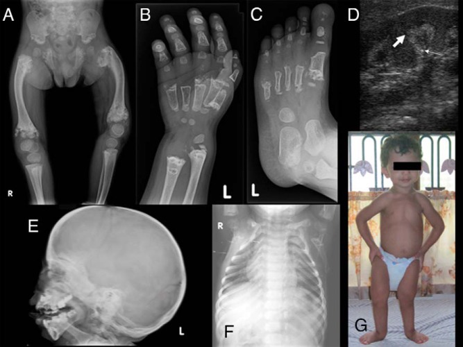
Objectives: Establishing the diagnosis of JMC during infancy or early childhood can be challenging, especially in the absence of family history and/or overt hypercalcemia. We therefore sought to provide radiographic findings supporting this diagnosis early in life.
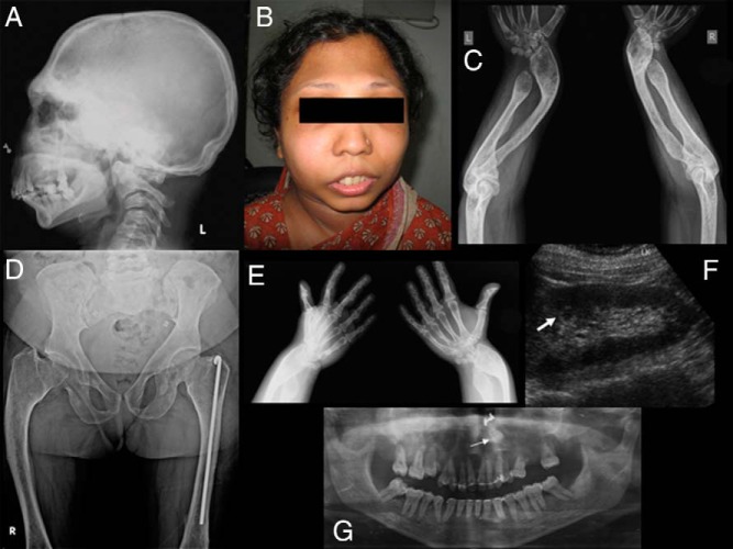
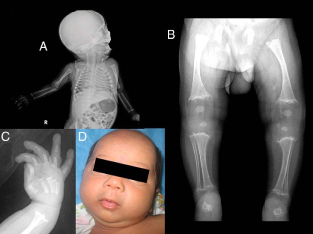
Patients and methods: Three patients, a mother and her two sons, had radiographic evidence for JMC. However, obvious hypercalcemia and suppressed PTH levels were encountered only in both affected children. Sanger sequencing and endonuclease (SphI) digestion of PCR-amplified genomic DNA were performed to search for the H223R-PTH1R mutation.
Results: The heterozygous H223R mutation was identified in all three affected individuals. Surprisingly, however, the now 38-year-old mother was never overtly hypercalcemic and was therefore not diagnosed until her sons were found to be affected by JMC at the ages of 28 months and 40 days, respectively. The presented radiographic findings at different ages will help diagnose other infants/toddlers suspected of having JMC.
Conclusion: The H223R mutation is typically associated with profound hypercalcemia despite low/normal PTH levels. However, the findings presented herein show that overt hypercalcemia is not always encountered in JMC, even if caused by this relatively frequent mutation, which is similar to observations with other PTH1R mutations that show less constitutive activity.
Figures
References
-
- Jansen M. Über atypische chondrodystrophie (achondroplasie) und über eine noch nicht beschriebene angeborene wachstumsstörung des knochensystems: metaphysäre dysostosis. Zeitschr Orthop Chir. 1934;61:253–286.
-
- Gram PB, Fleming JL, Frame B, Fine G. Metaphyseal chondrodysplasia of Jansen. J Bone Joint Surg. 1959;41A:951–959. - PubMed
-
- Jaffe HL. Certain other anomalies of skeletal development. In: Jaffe HL, ed. Metabolic, Degenerative, and Inflammatory Diseases of Bones and Joints. Philadelphia: Lea and Feibiger; 1972:222–226.
-
- Frame B, Poznanski AK. Conditions that may be confused with rickets. In: DeLuca HF, Anast CS, eds. Pediatric Diseases Related to Calcium. New York: Elsevier; 1980:269–289.
-
- Silve C, Jüppner H. Genetic disorders caused by mutations in the PTH/PTHrP receptor and down-stream effector molecules. In: Bilezikian J, ed. The Parathyroids: Basic and Clinical Concepts. San Diego: Academic Press; 2015:587–605.
Publication types
MeSH terms
Substances
Supplementary concepts
Grants and funding
LinkOut - more resources
Full Text Sources
Other Literature Sources
Research Materials
