Clinical Trial
. 2016 Jul 14;375(2):143-53.
doi: 10.1056/NEJMoa1601202.

Ipilimumab for Patients with Relapse after Allogeneic Transplantation

Affiliations
Clinical Trial

Ipilimumab for Patients with Relapse after Allogeneic Transplantation

Matthew S Davids et al. N Engl J Med. .

Abstract

Background: Loss of donor-mediated immune antitumor activity after allogeneic hematopoietic stem-cell transplantation (HSCT) permits relapse of hematologic cancers. We hypothesized that immune checkpoint blockade established by targeting cytotoxic T-lymphocyte-associated protein 4 with ipilimumab could restore antitumor reactivity through a graft-versus-tumor effect.

Methods: We conducted a phase 1/1b multicenter, investigator-initiated study to determine the safety and efficacy of ipilimumab in patients with relapsed hematologic cancer after allogeneic HSCT. Patients received induction therapy with ipilimumab at a dose of 3 or 10 mg per kilogram of body weight every 3 weeks for a total of 4 doses, with additional doses every 12 weeks for up to 60 weeks in patients who had a clinical benefit.

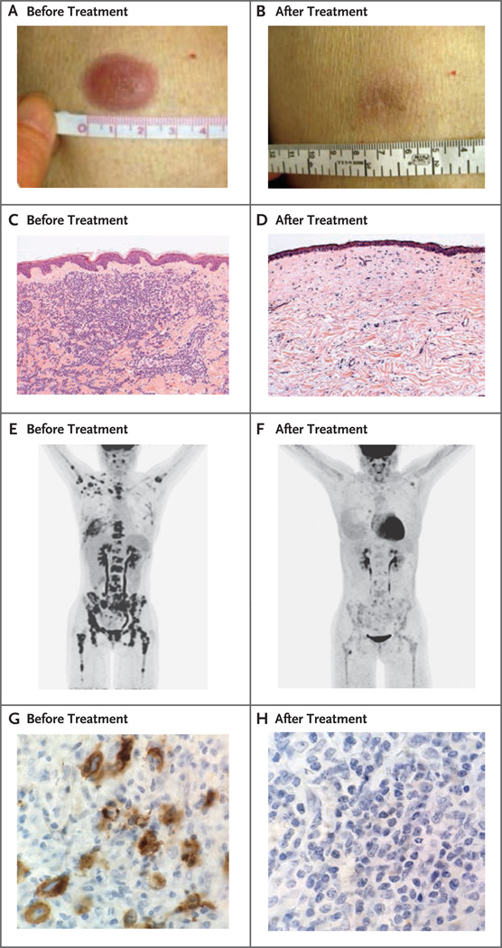
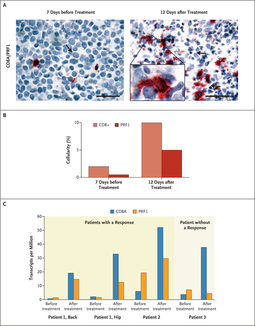
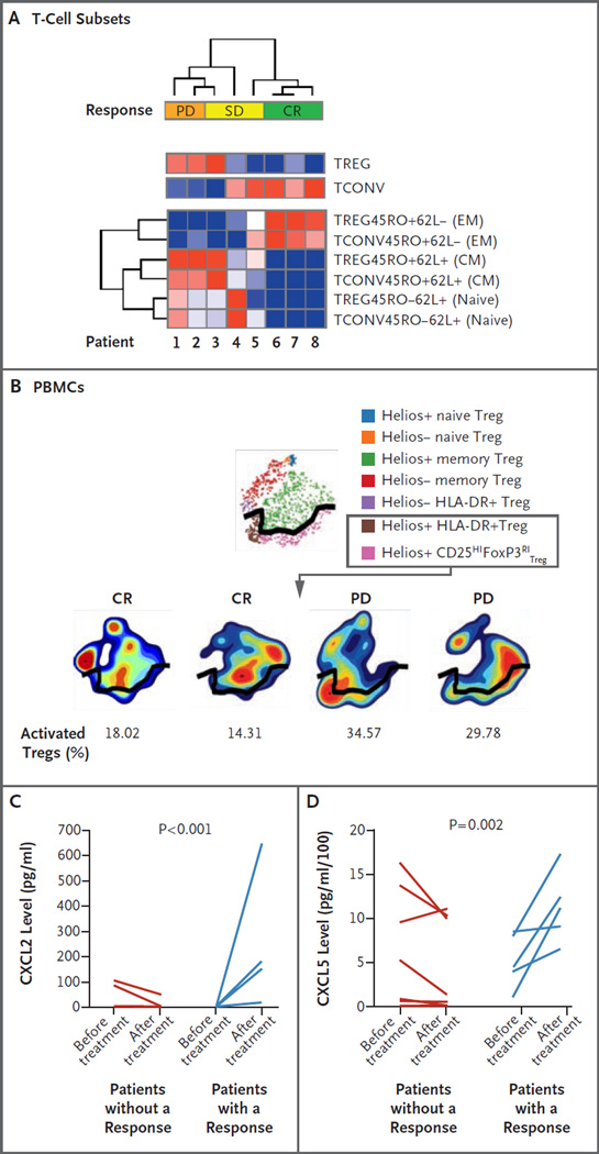
Results: A total of 28 patients were enrolled. Immune-related adverse events, including one death, were observed in 6 patients (21%), and graft-versus-host disease (GVHD) that precluded further administration of ipilimumab was observed in 4 patients (14%). No responses that met formal response criteria occurred in patients who received a dose of 3 mg per kilogram. Among 22 patients who received a dose of 10 mg per kilogram, 5 (23%) had a complete response, 2 (9%) had a partial response, and 6 (27%) had decreased tumor burden. Complete responses occurred in 4 patients with extramedullary acute myeloid leukemia and 1 patient with the myelodysplastic syndrome developing into acute myeloid leukemia. Four patients had a durable response for more than 1 year. Responses were associated with in situ infiltration of cytotoxic CD8+ T cells, decreased activation of regulatory T cells, and expansion of subpopulations of effector T cells in the blood.

Conclusions: Our early-phase data showed that administration of ipilimumab was feasible in patients with recurrent hematologic cancers after allogeneic HSCT, although immune-mediated toxic effects and GVHD occurred. Durable responses were observed in association with several histologic subtypes of these cancers, including extramedullary acute myeloid leukemia. (Funded by the National Institutes of Health and others; ClinicalTrials.gov number, NCT01822509.).


Figures

Figure 1
Figure 1. Clinical and Histopathological Responses to Ipilimumab in Patients with Leukemia Cutis and Hodgkin’s Lymphoma
Panel A shows a lesion in the skin of a patient with leukemia cutis before treatment, and Panel B shows the clinical response after treatment. Panel C shows sheets of neoplastic cells before treatment in the same patient, and Panel D shows the histopathological response after treatment, with fibrosis and sparse chronic inflammation without evidence of cancer (hematoxylin and eosin, low magnification in both panels). Panel E shows a positron-emission tomographic and computed tomographic (PET-CT) image before treatment and Panel F shows the response on PET-CT after treatment in a patient with Hodgkin’s lymphoma. Panel G shows a bone marrow biopsy specimen in the same patient before treatment, with staining for CD30 showing extensive malignant lymphocyte infiltration (high magnification), and Panel H shows a bone marrow biopsy specimen after treatment, with no evidence of cancer (high magnification).
Figure 2
Figure 2. Immunohistochemical and Transcriptional Analysis of In Situ Leukemic Responses to Ipilimumab
Panel A shows immunohistochemical images (at high magnification) of costaining for CD8+ T cells (red) and perforin (brown) before and after the initial dose of ipilimumab in a patient with leukemia cutis (Patient 1) who had a complete remission. The arrow on the left side of the panel indicates a leukemic blast; the arrows on the right side of the panel indicate cells undergoing apoptosis. The scale bars represent 50 µm. The inset (right) shows CD8+ T cells (red) contacting multiple leukemic cells with perforin staining (brown). Panel B shows quantitation by means of visual estimation for the stains in Panel A. Panel C shows gene-expression analyses of CD8A and PRF1 from biopsy specimens obtained before and after treatment with ipilimumab in two patients with leukemia who had a response and one patient who did not have a response. Biopsy specimens from two different sites of leukemia cutis (the back and hip) were obtained from Patient 1 before and after treatment.
Figure 3
Figure 3. Systemic Immunologic Correlative Studies in Patients Who Received Ipilimumab, According to Response Status
Panel A is a heat map showing the results of unsupervised clustering. These results are expressed as the percentage of T-cell subsets, as assessed by means of flow cytometry of peripheral-blood mononuclear cells (PBMCs) 8 weeks after administration of ipilimumab, in patients with progressive disease (PD), stable disease (SD), or a complete response (CR). The top two rows represent the percentage of total regulatory T-cells (Tregs) and conventional T-cells (Tconvs), and the bottom six rows represent T-cell subsets of these two populations, including Treg effector memory (EM) cells, Tconv EM cells, Treg central memory (CM) cells, Tconv CM cells, Treg naive cells, and Tconv naive cells. Minimum values (dark blue) and maximum values (dark red) in each row are shown. Plus signs indicate positive expression, and minus signs negative expression. Panel B shows a mass cytometric analysis of PBMCs at 8 weeks after administration of ipilimumab. Shown are activated Tregs (the population of cells below the black dividing line) in two patients who had a complete response as compared with two patients whose best response was PD. Cells expressing the Helios, HLA-DR, and CD25HIFoxP3RI markers are considered to be activated Tregs. Panel C shows levels of chemokine (C-X-C motif) ligand 2 (CXCL2), and Panel D shows levels of chemokine (C-X-C motif) ligand 5 (CXCL5) in the plasma of patients at baseline as compared with 8 weeks after treatment with ipilimumab, according to response status. Patients with a response included four patients with a CR and one with SD and some antitumor activity.

Comment in

References

    1. Antin JH. Graft-versus-leukemia: no longer an epiphenomenon. Blood. 1993;82:2273–2277. - PubMed
    1. Alyea EP, Kim HT, Ho V, et al. Comparative outcome of nonmyeloablative and myeloablative allogeneic hematopoietic cell transplantation for patients older than 50 years of age. Blood. 2005;105:1810–1814. - PubMed
    1. Mielcarek M, Storer BE, Flowers ME, Storb R, Sandmaier BM, Martin PJ. Outcomes among patients with recurrent high-risk hematologic malignancies after allogeneic hematopoietic cell transplantation. Biol Blood Marrow Transplant. 2007;13:1160–1168. - PubMed
    1. Thanarajasingam G, Kim HT, Cutler C, et al. Outcome and prognostic factors for patients who relapse after allogeneic hematopoietic stem cell transplantation. Biol Blood Marrow Transplant. 2013;19:1713–1718. - PMC - PubMed
    1. Soiffer RJ. Donor lymphocyte infusions for acute myeloid leukaemia. Best Pract Res Clin Haematol. 2008;21:455–466. - PubMed

Publication types

MeSH terms

Associated data