

Pinpointing a Mechanistic Switch Between Ketoreduction and "Ene" Reduction in Short-Chain Dehydrogenases/Reductases
- PMID: 27411040
- PMCID: PMC4988501
- DOI: 10.1002/anie.201603785
Pinpointing a Mechanistic Switch Between Ketoreduction and "Ene" Reduction in Short-Chain Dehydrogenases/Reductases
Abstract
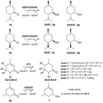
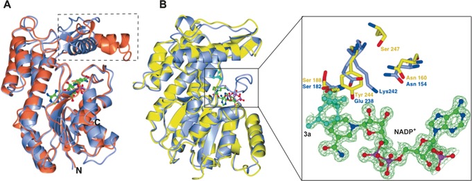
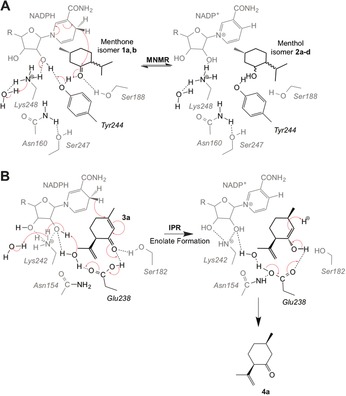
Three enzymes of the Mentha essential oil biosynthetic pathway are highly homologous, namely the ketoreductases (-)-menthone:(-)-menthol reductase and (-)-menthone:(+)-neomenthol reductase, and the "ene" reductase isopiperitenone reductase. We identified a rare catalytic residue substitution in the last two, and performed comparative crystal structure analyses and residue-swapping mutagenesis to investigate whether this determines the reaction outcome. The result was a complete loss of native activity and a switch between ene reduction and ketoreduction. This suggests the importance of a catalytic glutamate vs. tyrosine residue in determining the outcome of the reduction of α,β-unsaturated alkenes, due to the substrate occupying different binding conformations, and possibly also to the relative acidities of the two residues. This simple switch in mechanism by a single amino acid substitution could potentially generate a large number of de novo ene reductases.
Keywords: Mentha essential oil biosynthesis; biotransformations; isopiperitenone reductase; short-chain dehydrogenases/reductases; structure elucidation.
© 2016 The Authors. Published by Wiley-VCH Verlag GmbH & Co. KGaA.
Figures
References
-
- None
-
- Ringer K. L., McConkey M. E., Davis E. M., Rushing G. W., Croteau R., Arch. Biochem. Biophys. 2003, 418, 80–92; - PubMed
-
- Toogood H. S., Cheallaigh A. N., Tait S., Mansell D. J., Jervis A., Lygidakis A., Humphreys L., Takano E., Gardiner J. M., Scrutton N. S., ACS Synth. Biol. 2015, 4, 1112–1123. - PubMed
Publication types
MeSH terms
Substances
Grants and funding
LinkOut - more resources
Full Text Sources
Other Literature Sources
