. 2016 Jul 12;7(1):19-28.
doi: 10.1016/j.stemcr.2016.05.004.

Tissue-Engineered Vascular Rings from Human iPSC-Derived Smooth Muscle Cells

Affiliations

Tissue-Engineered Vascular Rings from Human iPSC-Derived Smooth Muscle Cells

Biraja C Dash et al. Stem Cell Reports. .

Abstract

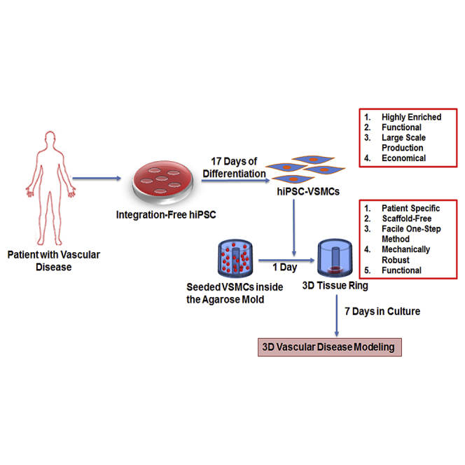
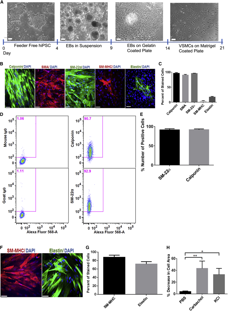
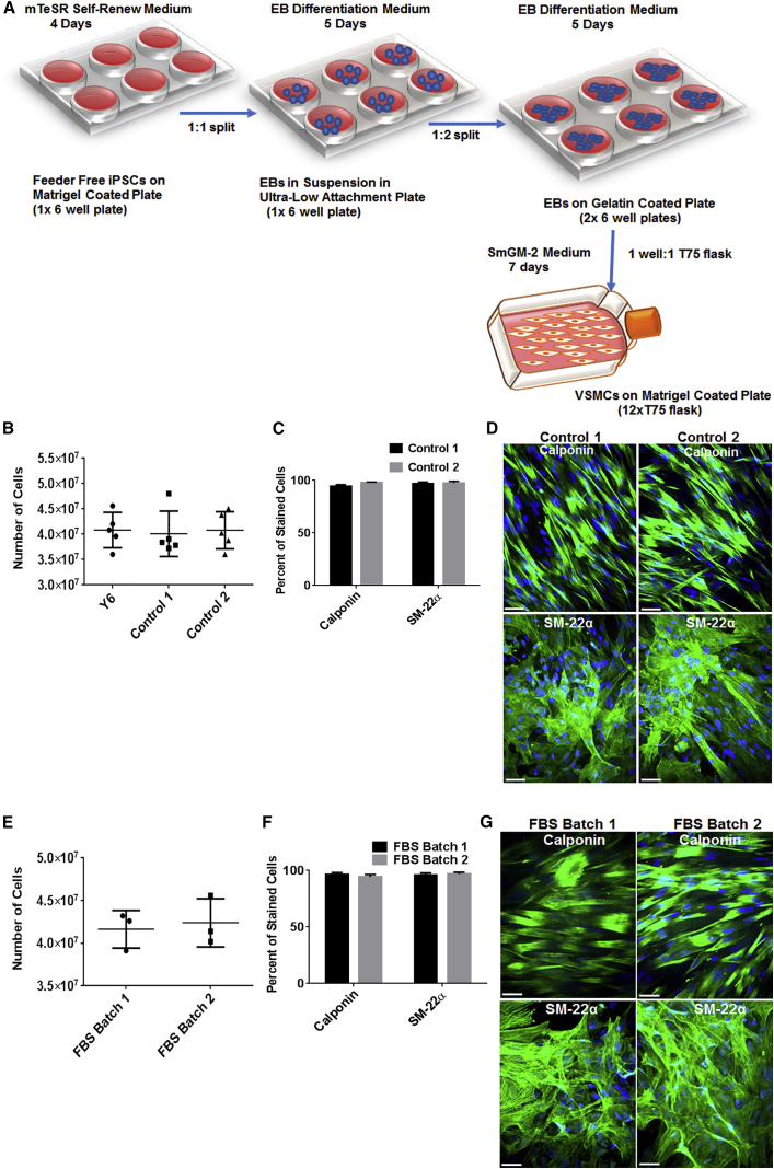
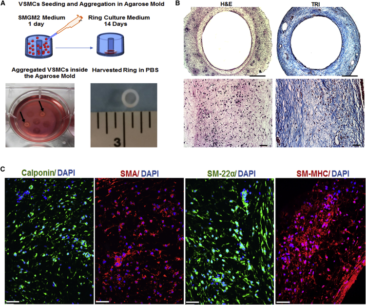
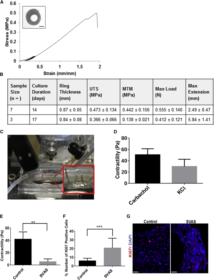
There is an urgent need for an efficient approach to obtain a large-scale and renewable source of functional human vascular smooth muscle cells (VSMCs) to establish robust, patient-specific tissue model systems for studying the pathogenesis of vascular disease, and for developing novel therapeutic interventions. Here, we have derived a large quantity of highly enriched functional VSMCs from human induced pluripotent stem cells (hiPSC-VSMCs). Furthermore, we have engineered 3D tissue rings from hiPSC-VSMCs using a facile one-step cellular self-assembly approach. The tissue rings are mechanically robust and can be used for vascular tissue engineering and disease modeling of supravalvular aortic stenosis syndrome. Our method may serve as a model system, extendable to study other vascular proliferative diseases for drug screening. Thus, this report describes an exciting platform technology with broad utility for manufacturing cell-based tissues and materials for various biomedical applications.


Figures

None
Graphical abstract
Figure 1
Figure 1
Characterization of hiPSC-VSMCs (A) Schematic diagram along with representative images showing the hiPSC-VSMC differentiation method. Scale bar represents 100 μm. (B) Immunofluorescence images of cells stained with smooth muscle cell markers calponin, SMA, SM-22α, SM-MHC, and elastin in hiPSC-VSMCs cultured in SmGM-2 medium for 7 days. Red (SMA and SM-MHC), green (SM-22α, calponin, and elastin), and blue (nuclei). Scale bar represents 50 μm. The Y6 hiPSC line was used to derive VSMCs. (C) Graph showing percentage of VSMCs positive for calponin, SMA, SM-22α, SM-MHC, and elastin in three independent experiments (mean ± SD; n = 3 independent experiments). (D) Fluorescence-activated cell sorting (FACS) data showing VSMC purity, quantified with smooth muscle cell-specific markers SM-22α and calponin. Goat and mouse IgGs were used as negative controls for SM-22α and calponin, respectively. (E) Graph showing percentage of VSMCs positive for SM-22α and calponin in three independent FACS experiments (mean ± SD; n = 3 independent experiments). (F) Immunofluorescence images of hiPSC-VSMCs cultured in a medium containing 0.5% FBS and 1 ng/ml TGF-β1 for 10 days and stained for SM-MHC and elastin. Red (SM-MHC), green (elastin), and blue (nuclei). Scale bar represents 50 μm. (G) Graph showing percentage of VSMCs positive for SM-MHC and elastin in three independent experiments. (H) Graph showing percent decrease in cell area in response to agonists KCl and carbachol. Unpaired Student’s t test was performed to determine the statistical difference between different groups (mean ± SD; n = 3 independent experiments, p < 0.05, ∗∗p < 0.01). See Figures S1–S3 and Tables S1 and S2 for additional controls and supporting information.
Figure 2
Figure 2
Larger-Scale VSMC Derivation from hiPSCs (A) Schematic diagram showing the procedure for derivation of hiPSC-VSMCs with high purity. (B–D) Characterization of hiPSC-VSMCs derived from two other hiPSC lines, control 1 and control 2. Control 1 and control 2 hiPSC-VSMC purity was quantified using calponin and SM-22α markers. (B) Resulting cell numbers from replicate derivations of VSMCs from Y6, control 1, and control 2 hiPSCs after 17 days of differentiation (n = 3 independent experiments). (C) Graph showing percentage of VSMCs positive for calponin or SM-22α in three independent experiments. (D) Representative immunofluorescence images of cells stained with calponin and SM-22α. Green stains for both calponin and SM-22. Blue stains for nuclei. Scale bar represents 50 μm. (E–G) Characterization of Y6-VSMCs derived using two different batches of FBS. (E) Quantification of cell numbers generated from hiPSC-VSMC derivations using two different batches of serum (n = 3 independent experiments). (F) Graph showing percentage of positive VSMCs for calponin and SM-22α in three independent experiments. (G) Immunofluorescence images of cells stained with calponin and SM-22α. Green stains for both calponin and SM-22α. Blue stains for nuclei. Scale bar represents 50 μm. The data are represented as means ± SD. Also see Figure S2.
Figure 3
Figure 3
Tissue Ring Fabrication and Characterization Using Y6 hiPSC-VSMCs (A) Schematic showing tissue ring fabrication by cellular self-assembly, and photographs of tissue rings in the agarose mold (black arrow) and a harvested tissue ring. (B) Histochemical analysis of the tissue rings with H&E (left) and Masson's trichrome (TRI: right) staining. The pink in H&E staining represents cytoplasm and the blue in Masson's trichrome represents collagen. The dark purple in both H&E and Masson's trichrome staining represents nuclei. Scale bars represent 500 and 100 μm, respectively, for lower and higher magnification images. (C) Immunofluorescence analysis of calponin (green), SMA (red), SM-22α (green), and SM-MHC (red) present in the tissue rings after 14 days in culture. Blue stains for nuclei. Scale bar represents 50 μm. Also see Figure S4.
Figure 4
Figure 4
Mechanical and Functional Characterization of hiPSC-VSMC Rings (A) Representative stress-strain curve of a 14-day-old tissue ring (inset, digital image of ring used to measure thickness and calculate cross-sectional area; scale = 1 mm). (B) Results of mechanical tests from two independent batches of hiPSC-VSMC tissue rings (mean ± SD; n = 7 and 3 independent measurements for day 14 and 17, respectively). (C) Photograph showing VSMC tissue ring hooked on two micromanipulators and immersed in a temperature-controlled perfusion bath containing Tyrode's solution for the contractility assay. (D) Graph showing change in contractility (Pascal) in response to the agonists KCl and carbachol. PBS was used as negative control. (E) Graph showing change in contractility (Pascal) of control and SVAS rings in response to carbachol. Unpaired Student’s t test was performed to determine statistical difference between different groups (mean ± SD; n = 3 independent experiments, ∗∗p < 0.01). PBS was used as negative control. (F) Graph showing percent number of Ki67-positive cells in control and SVAS rings. The data are represented as mean ± SD. Unpaired Student’s t test was performed to determine statistical difference between different groups (n = 3 independent experiments, ∗∗∗p < 0.001). (G) Representative immunofluorescence images of control and SVAS rings stained with Ki67. Red (Ki67) and blue (nuclei). Scale bar represents 50 μm. Also see Figure S4.

Similar articles

Cited by

References

    1. Bajpai V.K., Mistriotis P., Loh Y.H., Daley G.Q., Andreadis S.T. Functional vascular smooth muscle cells derived from human induced pluripotent stem cells via mesenchymal stem cell intermediates. Cardiovasc. Res. 2012;96:391–400. - PMC - PubMed
    1. Chang S., Song S., Lee J., Yoon J., Park J., Choi S., Park J.K., Choi K., Choi C. Phenotypic modulation of primary vascular smooth muscle cells by short-term culture on micropatterned substrate. PLoS One. 2014;9:e88089. - PMC - PubMed
    1. Chen I.P., Fukuda K., Fusaki N., Iida A., Hasegawa M., Lichtler A., Reichenberger E.J. Induced pluripotent stem cell reprogramming by integration-free Sendai virus vectors from peripheral blood of patients with craniometaphyseal dysplasia. Cell Reprogram. 2013;15:503–513. - PMC - PubMed
    1. Cheung C., Bernardo A.S., Trotter M.W., Pedersen R.A., Sinha S. Generation of human vascular smooth muscle subtypes provides insight into embryological origin-dependent disease susceptibility. Nat. Biotechnol. 2012;30:165–173. - PMC - PubMed
    1. Dash B.C., Jiang Z., Suh C., Qyang Y. Induced pluripotent stem cell-derived vascular smooth muscle cells: methods and application. Biochem. J. 2015;465:185–194. - PMC - PubMed

Publication types

MeSH terms