Case Reports
. 2016 Jul 13;4(1):73.
doi: 10.1186/s40478-016-0342-3.

BK virus encephalopathy and sclerosing vasculopathy in a patient with hypohidrotic ectodermal dysplasia and immunodeficiency

Affiliations
Case Reports

BK virus encephalopathy and sclerosing vasculopathy in a patient with hypohidrotic ectodermal dysplasia and immunodeficiency

Armine Darbinyan et al. Acta Neuropathol Commun. .

Abstract

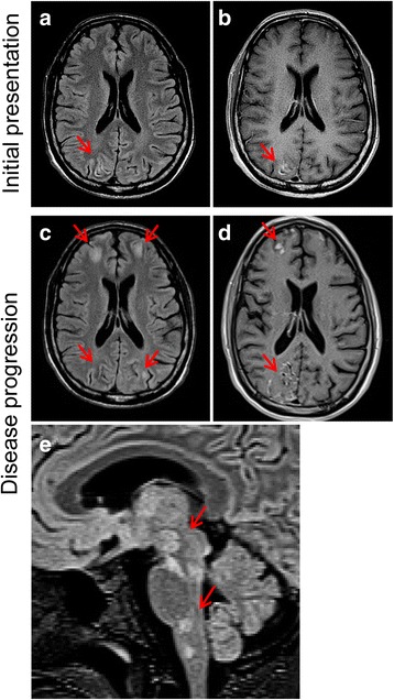
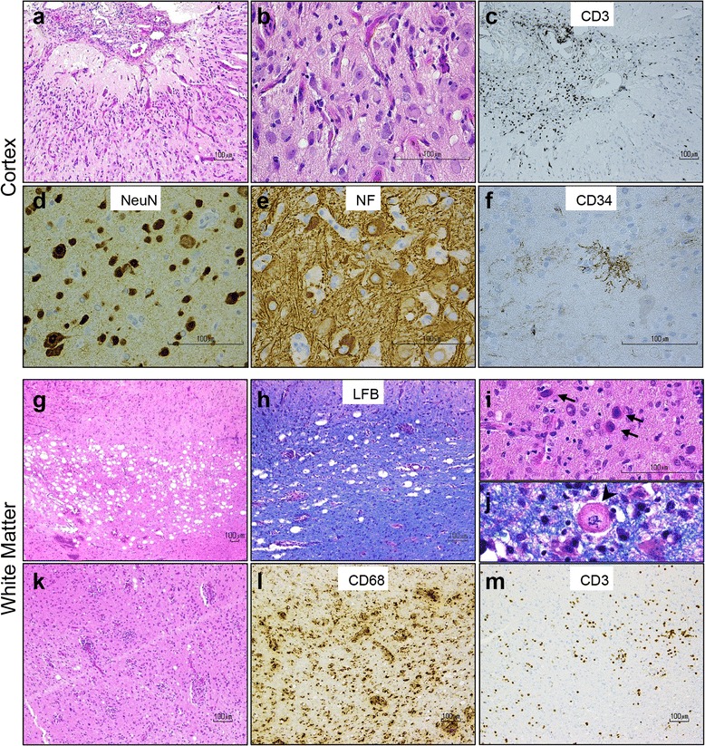
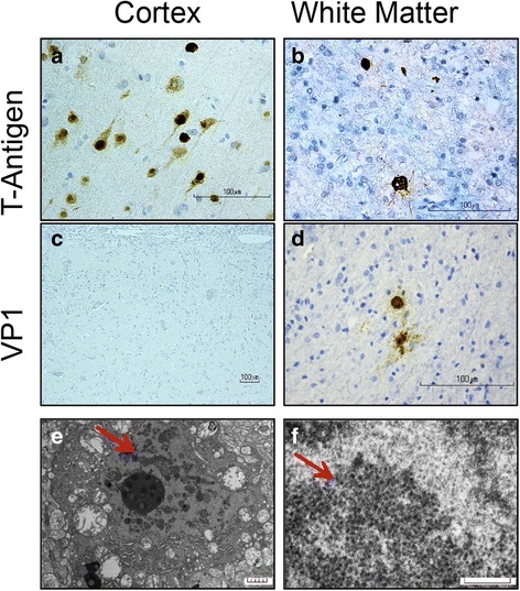
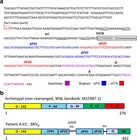
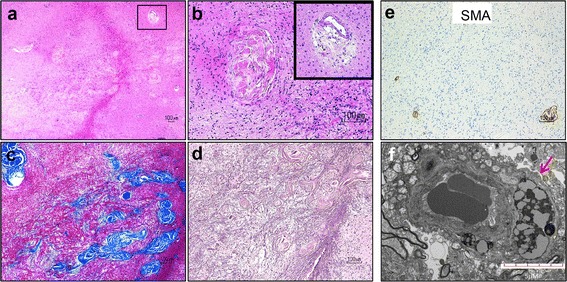
Human BK polyomavirus (BKV) is reactivated under conditions of immunosuppression leading most commonly to nephropathy or cystitis; its tropism for the brain is rare and poorly understood. We present a unique case of BKV-associated encephalopathy in a man with hypohidrotic ectodermal dysplasia and immunodeficiency (HED-ID) due to IKK-gamma (NEMO) mutation, who developed progressive neurological symptoms. Brain biopsy demonstrated polyomavirus infection of gray and white matter, with predominant involvement of cortex and distinct neuronal tropism, in addition to limited demyelination and oligodendroglial inclusions. Immunohistochemistry demonstrated polyoma T-antigen in neurons and glia, but expression of VP1 capsid protein only in glia. PCR analysis on both brain biopsy tissue and cerebrospinal fluid detected high levels of BKV DNA. Sequencing studies further identified novel BKV variant and disclosed unique rearrangements in the noncoding control region of the viral DNA (BKVN NCCR). Neuropathological analysis also demonstrated an unusual form of obliterative fibrosing vasculopathy in the subcortical white matter with abnormal lysosomal accumulations, possibly related to the patient's underlying ectodermal dysplasia. Our report provides the first neuropathological description of HED-ID due to NEMO mutation, and expands the diversity of neurological presentations of BKV infection in brain, underscoring the importance of its consideration in immunodeficient patients with unexplained encephalopathy. We also document novel BKVN NCCR rearrangements that may be associated with the unique neuronal tropism in this patient.

Keywords: BK virus; Ectodermal dysplasia; Encephalopathy; Fibrosing vasculopathy; HED-ID; IKK-gamma; NF-kappa-B essential modulator (NEMO); Polyomavirus.


Figures

Fig. 1
Fig. 1
Serial axial T2 FLAIR MRI (a, c) and corresponding axial contrast-enhanced T1 MRI (b, d) imaging of patient during initial presentation and after three months of disease progression when biopsy was performed. a Initial MRI (Day 1 of hospitalization) shows cortical gray matter with increased T2 FLAIR signal that is most prominent in the right occipital lobe (arrow) and just appreciable in the left occipital lobe. The related sulci are prominent, not compressed. The juxta-cortical white matter appears normal. b There is prominent contrast enhancement of portions of the affected cortices with no leptomeningeal enhancement. c-d Surveillance axial T2 FLAIR MRI (c, Day 90 of hospitalization,) and contrast-enhanced T1 MRI (d, Day 82 of hospitalization,) show disease progression with prominent involvement of both occipital and both frontal lobes. e T2 MRI through the deep gray matter and brain stem (sagittal) on Day 81 of hospitalization demonstrates patchy regions with signal abnormality in the basal ganglia, thalami and brain stem, including the medial left thalamus, right lateral geniculate nucleus, inferior colliculus, interpeduncular nuclei, dorsal pontine nuclei, and lateral medullary nuclei
Fig. 2
Fig. 2
Histological analysis of right occipital brain biopsy reveals unusual form of inflammatory encephalopathy. a-b Hematoxylin and eosin (H&E) stains show chronic meningitis and neocortex with architectural neuronal disorganization, associated with reactive vasculature, gliosis, microglial activation, and mild chronic inflammation. c CD3+ T-lymphocytes predominate in the meninges. d Many cortical neurons display dysmorphic features, such as increased size, displaced Nissl substance, clustering, and e abnormal phosphorylated neurofilament accumulation in their cell body. f Tuft-like ramified CD34-positive processes are also seen in the neocortex. g-j The subcortical white matter contains multifocal vacuolization associated with minimal myelin loss on luxol fast blue (LFB) stain, and scattered bizarre oligodendroglial-like cells with enlarged nuclei containing glassy, homogeneous nuclear inclusions (i, arrows) and Creutzfeldt-like astrocytes (j, arrowhead). k-m Prominent chronic perivascular and intraparenchymal inflammatory infiltrate in the white matter is composed of CD68+ macrophage/microglia and CD3+ T-lymphocytes
Fig. 3
Fig. 3
Polyomavirus infection in cortex and white matter by immunohistochemistry and electron microscopy. a-b Immunostaining with the cross-reacting polyomavirus antibody anti-SV40 T-Antigen shows strong nuclear positivity in cortical neurons, predominantly in layers II-V (a) and in scattered enlarged oligodendroglial and astrocytic nuclei (b) in the white matter. c-d Immunoreactivity with the cross-reacting antibody anti-VP1 capsid protein is absent in cortical neurons (c) but is present in scattered cells with glial appearance in the white matter (d). e Electron micrographs reveal a neural cell with aggregates of spherical particles admixed with chromatin (arrow), scale bar = 5 μm. f At higher power, 30–40 nm in diameter non-enveloped icosahedral viral particles are observed, focally forming sheets and paracrystalline arrays (arrow); scale bar = 0.26 μm
Fig. 4
Fig. 4
Sequencing confirms BK virus infection in brain tissue and identifies unique BKVN NCCR region. a Sequence analysis of BKVN NCCR region. Start codon for the late coding region is shown by red arrow. The origin of viral replication (ori) and TATA box are depicted. T-Ag binding sites are underlined. The BKVN P blocks appear in two variants consisting of 41 (ΔP41) and 25 (ΔP26) nucleotides are shown in blue or red, respectively. Each of these blocks is similar to the initial portion of P block of archetype. Insertions/variations are also highlighted (pink). b Schematic comparison of NCCR sequence between BKV archetypal and BKVN. The NCCR region of the archetypal BKV contains five sequence blocks denoted O-143 (with ori), P-68, Q-39, R-63 and S-63 where the numbers indicate base pairs. The BKVN NCCR contains block O-143, two tandem repeats composed of portions of block P (ΔP41 and ΔP26) and separated by polyA stretch. Blocks Q and R follow the second ΔP26
Fig. 5
Fig. 5
Unusual form of sclerosing vasculopathy in HED-ID. a-b Hematoxylin and eosin stain shows vessels in the white matter with variably obliterated lumina, hyalinization, and perivascular hemosiderin deposits (inset in b). c-d Concentric lamellated pattern of collagen accumulation is seen on trichrome (c), extending into the adjacent brain parenchyma and forming a reticulin-rich scar (d). e Sclerotic vessels lack smooth muscle fibers in their walls. f EM shows capillary vessel with aberrant accumulation of lysosomes (arrow); scale bar = 5 μm

Similar articles

Cited by

References

    1. Abend JR, Joseph AE, Das D, Campbell-Cecen DB, Imperiale MJ. A truncated T antigen expressed from an alternatively spliced BK virus early mRNA. J Gen Virol. 2009;90:1238–1245. doi: 10.1099/vir.0.009159-0. - DOI - PMC - PubMed
    1. Aradhya S, Courtois G, Rajkovic A, Lewis RA, Levy M, Israel A, Nelson DL. Atypical forms of incontinentia pigmenti in male individuals result from mutations of a cytosine tract in exon 10 of NEMO (IKK-gamma) Am J Hum Genet. 2001;68:765–771. doi: 10.1086/318806. - DOI - PMC - PubMed
    1. Azzi A, Cesaro S, Laszlo D, Zakrzewska K, Ciappi S, De Santis R, Fanci R, Pesavento G, Calore E, Bosi A. Human polyomavirus BK (BKV) load and haemorrhagic cystitis in bone marrow transplantation patients. J Clin Virol. 1999;14:79–86. doi: 10.1016/S1386-6532(99)00055-4. - DOI - PubMed
    1. Barcena-Panero A, Echevarria JE, Van Ghelue M, Fedele G, Royuela E, Gerits N, Moens U. BK polyomavirus with archetypal and rearranged non-coding control regions is present in cerebrospinal fluids from patients with neurological complications. J Gen Virol. 2012;93:1780–1794. doi: 10.1099/vir.0.042143-0. - DOI - PubMed
    1. Barcena-Panero A, Van Ghelue M, Khan MT, Echevarria JE, Fedele G, Moens U. BK virus-associated infection in cerebrospinal fluid of neurological patients and mutation analysis of the complete VP1 gene in different patient groups. J Cell Physiol. 2012;227:136–145. doi: 10.1002/jcp.22711. - DOI - PubMed

Publication types

MeSH terms

Substances