. 2016 Dec;21(3-4):81-88.
doi: 10.1007/s10911-016-9358-3. Epub 2016 Jul 13.

p120-Catenin Is Critical for the Development of Invasive Lobular Carcinoma in Mice

Affiliations

p120-Catenin Is Critical for the Development of Invasive Lobular Carcinoma in Mice

Milou Tenhagen et al. J Mammary Gland Biol Neoplasia. 2016 Dec.

Abstract

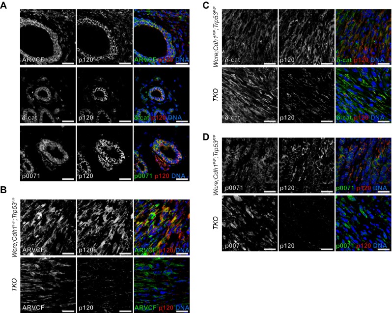
Loss of E-cadherin expression is causal to the development of invasive lobular breast carcinoma (ILC). E-cadherin loss leads to dismantling of the adherens junction and subsequent translocation of p120-catenin (p120) to the cytosol and nucleus. Although p120 is critical for the metastatic potential of ILC through the regulation of Rock-dependent anoikis resistance, it remains unknown whether p120 also contributes to ILC development. Using genetically engineered mouse models with mammary gland-specific inactivation of E-cadherin, p120 and p53, we demonstrate that ILC formation induced by E-cadherin and p53 loss is severely impaired upon concomitant inactivation of p120. Tumors that developed in the triple-knockout mice were mostly basal sarcomatoid carcinomas that displayed overt nuclear atypia and multinucleation. In line with the strong reduction in ILC incidence in triple-knockout mice compared to E-cadherin and p53 double-knockout mice, no functional redundancy of p120 family members was observed in mouse ILC development, as expression and localization of ARVCF, p0071 or δ-catenin was unaltered in ILCs from triple-knockout mice. In conclusion, we show that loss of p120 in the context of the p53-deficient mouse models is dominant over E-cadherin inactivation and its inactivation promotes the development of basal, epithelial-to-mesenchymal-transition (EMT)-type invasive mammary tumors.

Keywords: Breast cancer; Invasive lobular carcinoma; Mouse model; p120.


Conflict of interest statement

Compliance with Ethical Standards Grants Research was supported by grants from the Netherlands Organization for Scientific Research (NWO/ZonMW-VIDI 016.096.318), Foundation Vrienden UMC Utrecht (11.081) and the Dutch Cancer Society (KWF-UU-2011-5230). Conflict of Interest The authors declare that they have no conflict of interest.

Figures

Fig. 1
Fig. 1
Tumor incidence in Wcre females carrying conditional Ctnnd1, Cdh1 and Trp53 alleles. Kaplan-Meier tumor-free survival curves are shown for mammary tumors from Wcre;Cdh1 F/F;Trp53 F/F (red curve) TKO (black curve) and Wcre;Ctnnd1 F/+;Cdh1 F/F;Trp53 F/F (green curve). Mice were sacrificed when tumors reached an average diameter of 10 mm
Fig. 2
Fig. 2
E-cadherin and p120 expression in the mammary tumor spectrum. a-b Sections showing representative examples for mouse ILC (mILC) (a) and solid carcinoma/carcinosarcoma (SC/CS) (b) for the different genotypes studied. Sections were stained for E-cadherin (middle panels) and p120 (bottom panels). In TKO tumors p120 is only expressed by stromal cells (arrows). Nuclear atypia is present in p120 negative tumors (arrow heads). The middle and bottom panels are magnifications that correspond to the area indicated in the upper panels. Size bar =25 μm. c Distribution of AC (green), mILC (blue) and SC/CS tumors (orange) in mammary glands of Wcre;Cdh1 F/F;Trp53 F/F, Wcre;Ctnnd1 F/+;Cdh1 F/F;Trp53 F/F and TKO female mice
Fig. 3
Fig. 3
Metastases of mouse tumors from the TKO mouse model. Tumor dissemination showing metastasis into the axillary lymph node (a), lungs (b), liver (c) and spleen (d). Primary tumors were diagnosed as carcinosarcoma. Dotted lines outline the metastatic area (M). The bottom panels are magnifications that correspond to the area indicated in the top panels. Size bar =25 μm
Fig. 4
Fig. 4
Comparative immunohistochemistry of mammary tumors. Analysis of marker expression in tumor types of Wcre;Cdh1 F/F;Trp53 F/F and TKO mice. Luminal cells were identified by CK8 expression, while CK14 staining was performed to detect basal cells. Vimentin (Vim) was used as a mesenchymal marker. The bottom panels are magnifications that correspond to the area indicated in the top panels. Size bar =50 μm
Fig. 5
Fig. 5
Expression of p120 family members in mILC. a p120 family member expression (left panels, green) in the mouse mammary gland. Expression of p120 is depicted in the middle panels (red). b-d Immunofluorescence showing expression of p120 (red) and its family members (green) ARVCF (b), δ-catenin (c) and p0071 (d) in mILCs from Wcre;Cdh1 F/F;Trp53 F/F (top panels) and TKO mice (bottom panels). DAPI (blue) was used to visualize nuclei. The merged images are shown in the right panels. Size bar =25 μm
Fig. 6
Fig. 6
Inactivation of E-cadherin or p120 has divergent consequences on mammary tumor etiology. WAP-cre dependent stochastic loss of p53 predisposes tumor-initiating cells to the development of non-metastatic luminal adenocarcinoma (AC) and basal solid carcinoma/carcinosarcoma (SC/CS) [11]. Inactivation of E-cadherin in this context drives the luminal tumor spectrum towards metastatic invasive lobular cancer (ILC). In contrast, p120 ablation tilts the balance towards formation of basal EMT-type SC/CS. In general, conditional loss of either E-cadherin or p120 results in invasive and metastatic tumors. Inactivation of the adherens junction through p120 loss is dominant over E-cadherin loss in TKO mice, largely preventing ILC development, and driving the formation of EMT-type basal mammary tumors. Arrows indicate mammary-specific Cre-LoxP mediated gene inactivation

References

    1. Jeanes A, Gottardi CJ, Yap AS. Cadherins and cancer: how does cadherin dysfunction promote tumor progression? Oncogene. 2008;27:6920–6929. doi: 10.1038/onc.2008.343. - DOI - PMC - PubMed
    1. Christgen M, Derksen PW. Lobular breast cancer: molecular basis, mouse and cellular models. Breast Cancer Res. 2015;17:1–9. doi: 10.1186/s13058-015-0517-z. - DOI - PMC - PubMed
    1. Vos CB, Cleton-Jansen AM, Berx G, de Leeuw WJ, ter Haar NT, van Roy F, et al. E-cadherin inactivation in lobular carcinoma in situ of the breast: an early event in tumorigenesis. Br J Cancer. 1997;76:1131–1133. doi: 10.1038/bjc.1997.523. - DOI - PMC - PubMed
    1. Berx G, Nollet F, De LWJF, Vijver MJ, De V, Cornelisse C, Van RF. E-cadherin as a tumour/invasion suppressor gene mutated in human lobular breast cancers. EMBO J. 1995;14:6107–6115. - PMC - PubMed
    1. Sarrió D, Pérez-Mies B, Hardisson D, Moreno-Bueno G, Suárez A, Cano A, et al. Cytoplasmic localization of p120ctn and E-cadherin loss characterize lobular breast carcinoma from preinvasive to metastatic lesions. Oncogene. 2004;23:3272–3283. doi: 10.1038/sj.onc.1207439. - DOI - PubMed

Publication types

LinkOut - more resources