

Quantitative Persulfide Site Identification (qPerS-SID) Reveals Protein Targets of H2S Releasing Donors in Mammalian Cells
- PMID: 27411966
- PMCID: PMC4944133
- DOI: 10.1038/srep29808
Quantitative Persulfide Site Identification (qPerS-SID) Reveals Protein Targets of H2S Releasing Donors in Mammalian Cells
Abstract
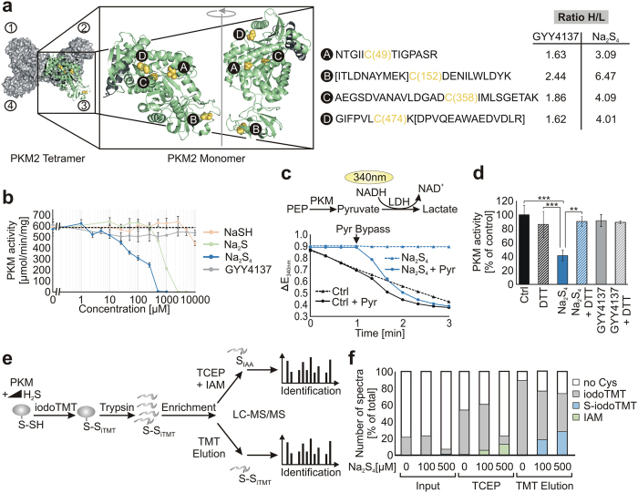
H2S is an important signalling molecule involved in diverse biological processes. It mediates the formation of cysteine persulfides (R-S-SH), which affect the activity of target proteins. Like thiols, persulfides show reactivity towards electrophiles and behave similarly to other cysteine modifications in a biotin switch assay. In this manuscript, we report on qPerS-SID a mass spectrometry-based method allowing the isolation of persulfide containing peptides in the mammalian proteome. With this method, we demonstrated that H2S donors differ in their efficacy to induce persulfides in HEK293 cells. Furthermore, data analysis revealed that persulfide formation affects all subcellular compartments and various cellular processes. Negatively charged amino acids appeared more frequently adjacent to cysteines forming persulfides. We confirmed our proteomic data using pyruvate kinase M2 as a model protein and showed that several cysteine residues are prone to persulfide formation finally leading to its inactivation. Taken together, the site-specific identification of persulfides on a proteome scale can help to identify target proteins involved in H2S signalling and enlightens the biology of H2S and its releasing agents.
Figures
Similar articles
-
Reaction of Hydrogen Sulfide with Disulfide and Sulfenic Acid to Form the Strongly Nucleophilic Persulfide.J Biol Chem. 2015 Nov 6;290(45):26866-26880. doi: 10.1074/jbc.M115.672816. Epub 2015 Aug 12. J Biol Chem. 2015. PMID: 26269587 Free PMC article.
-
Kinetics of formation and reactivity of the persulfide in the one-cysteine peroxiredoxin from Mycobacterium tuberculosis.J Biol Chem. 2019 Sep 13;294(37):13593-13605. doi: 10.1074/jbc.RA119.008883. Epub 2019 Jul 16. J Biol Chem. 2019. PMID: 31311857 Free PMC article.
-
Cysteine persulfides and polysulfides produced by exchange reactions with H2S protect SH-SY5Y cells from methylglyoxal-induced toxicity through Nrf2 activation.Redox Biol. 2017 Aug;12:530-539. doi: 10.1016/j.redox.2017.03.020. Epub 2017 Mar 24. Redox Biol. 2017. PMID: 28371750 Free PMC article.
-
Biological chemistry of hydrogen sulfide and persulfides.Arch Biochem Biophys. 2017 Mar 1;617:9-25. doi: 10.1016/j.abb.2016.09.018. Epub 2016 Sep 30. Arch Biochem Biophys. 2017. PMID: 27697462 Review.
-
Persulfides, at the crossroads between hydrogen sulfide and thiols.Essays Biochem. 2020 Feb 17;64(1):155-168. doi: 10.1042/EBC20190049. Essays Biochem. 2020. PMID: 32016341 Review.
Cited by
-
Biological hydropersulfides and related polysulfides - a new concept and perspective in redox biology.FEBS Lett. 2018 Jun;592(12):2140-2152. doi: 10.1002/1873-3468.13090. Epub 2018 May 24. FEBS Lett. 2018. PMID: 29754415 Free PMC article. Review.
-
N-Acetyl Cysteine Functions as a Fast-Acting Antioxidant by Triggering Intracellular H2S and Sulfane Sulfur Production.Cell Chem Biol. 2018 Apr 19;25(4):447-459.e4. doi: 10.1016/j.chembiol.2018.01.011. Epub 2018 Feb 8. Cell Chem Biol. 2018. PMID: 29429900 Free PMC article.
-
Cysteine residues in mitochondrial intermembrane space proteins: more than just import.Br J Pharmacol. 2019 Feb;176(4):514-531. doi: 10.1111/bph.14480. Epub 2018 Sep 28. Br J Pharmacol. 2019. PMID: 30129023 Free PMC article. Review.
-
Evidence of endogenously produced hydrogen sulfide (H2S) and persulfidation in male reproduction.Sci Rep. 2022 Jul 6;12(1):11426. doi: 10.1038/s41598-022-15360-x. Sci Rep. 2022. PMID: 35794129 Free PMC article.
-
Redox Signaling by Reactive Electrophiles and Oxidants.Chem Rev. 2018 Sep 26;118(18):8798-8888. doi: 10.1021/acs.chemrev.7b00698. Epub 2018 Aug 27. Chem Rev. 2018. PMID: 30148624 Free PMC article. Review.
References
-
- Bełtowski J. Hydrogen sulfide in pharmacology and medicine - An update. Pharmacol. Rep. 67, 647–658 (2015). - PubMed
-
- Li L. et al.. Characterization of a novel, water-soluble hydrogen sulfide-releasing molecule (GYY4137): New insights into the biology of hydrogen sulfide. Circulation 117, 2351–2360 (2008). - PubMed
Publication types
MeSH terms
Substances
LinkOut - more resources
Full Text Sources
Other Literature Sources
