. 2016 Jul 13;47(1):70.
doi: 10.1186/s13567-016-0354-9.

Productive replication of nephropathogenic infectious bronchitis virus in peripheral blood monocytic cells, a strategy for viral dissemination and kidney infection in chickens

Affiliations

Productive replication of nephropathogenic infectious bronchitis virus in peripheral blood monocytic cells, a strategy for viral dissemination and kidney infection in chickens

Vishwanatha R A P Reddy et al. Vet Res. .

Abstract

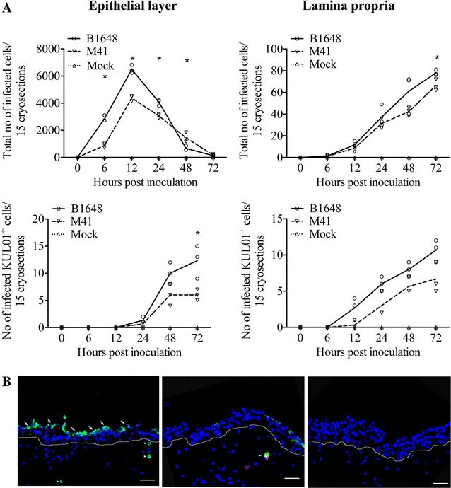
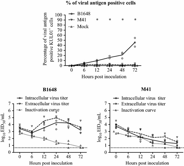
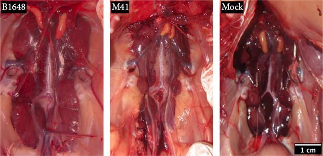
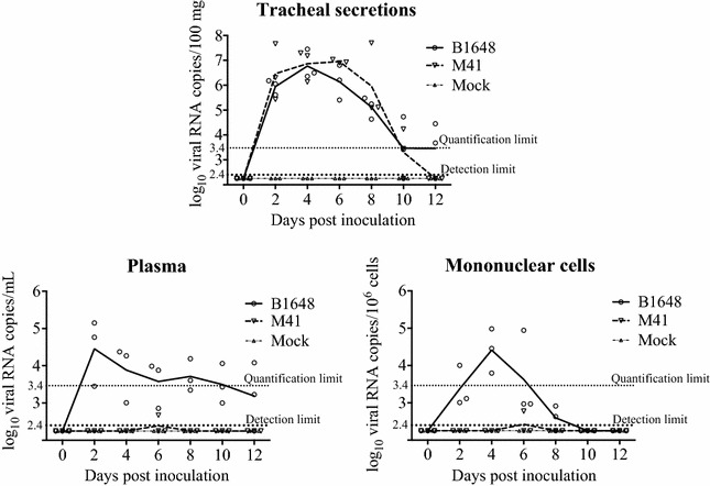
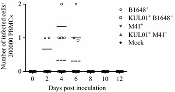
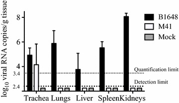
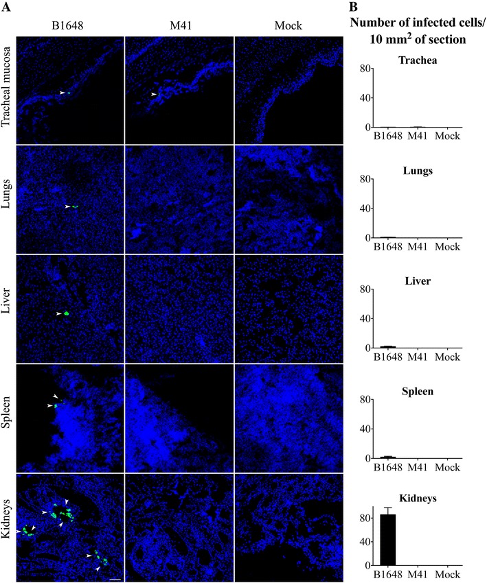
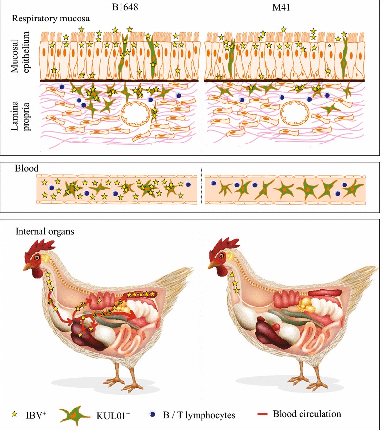
In the present study, the replication kinetics of nephropathogenic (B1648) and respiratory (Massachusetts-M41) IBV strains were compared in vitro in respiratory mucosa explants and blood monocytes (KUL01(+) cells), and in vivo in chickens to understand why some IBV strains have a kidney tropism. B1648 was replicating somewhat better than M41 in the epithelium of the respiratory mucosa explants and used more KUL01(+) cells to penetrate the deeper layers of the respiratory tract. B1648 was productively replicating in KUL01(+) monocytic cells in contrast with M41. In B1648 inoculated animals, 10(2.7-6.8) viral RNA copies/100 mg were detected in tracheal secretions at 2, 4, 6, 8, 10 and 12 days post inoculation (dpi), 10(2.4-4.5) viral RNA copies/mL in plasma at 2, 4, 6, 8, 10 and 12 dpi and 10(1.8-4.4) viral RNA copies/10(6) mononuclear cells in blood at 2, 4, 6 and 8 dpi. In M41 inoculated animals, 10(2.6-7.0) viral RNA copies/100 mg were detected in tracheal secretions at 2, 4, 6, 8 and 10 dpi, but viral RNA was not demonstrated in plasma and mononuclear cells (except in one chicken at 6 dpi). Infectious virus was detected only in plasma and mononuclear cells of the B1648 group. At euthanasia (12 dpi), viral RNA and antigen positive cells were detected in lungs, liver, spleen and kidneys of only the B1648 group and in tracheas of both the B1648 and M41 group. In conclusion, only B1648 can easily disseminate to internal organs via a cell-free and -associated viremia with KUL01(+) cells as important carrier cells.


Figures

Figure 1
Figure 1
IBV infected cell quantification (A) and antigen expression (B) in tracheal mucosa explants. A Number of IBV B1648 and M41 infected cells and infected KUL01+ cells were quantified in the epithelial layer and lamina propria of tracheal mucosa explants. Fifteen consecutive cryosections were analysed at 0, 6, 12, 24, 48 and 72 hpi to quantify infected cells. The number of infected KUL01+cells starts to increase from 12 hpi, when the total number of IBV infected cells starts to decrease. Lines represent the evolution of arithmetic means without any transformation of original data. Solid line represents mean values of B1648, dashed line represents mean values of M41 and dotted line represents mean values of mock. An asterisk (*) indicates a significant difference (P < 0.05) between B1648 and M41. B Representative confocal photomicrograph illustrating IBV infected cells (left panel) in the epithelium (arrows) and lamina propria (arrowheads), and IBV infected KUL01+cell (middle panel) in lamina propria (arrowhead) of the tracheal mucosa. Mock-inoculated tracheal mucosa (right panel). Green fluorescence visualises IBV antigens. KUL01+ cells are visualised by red fluorescence. Cell nuclei were stained with Hoechst (blue). White dotted line indicates the BM. Scale bar represents 50 μm.
Figure 2
Figure 2
IBV replication kinetics in blood monocytes (KUL01 + cells). The monocytes were inoculated with IBV B1648 or M41 at a m.o.i. = 5. Percentage of viral antigen positive KUL01+ cells was determined in B1648 and M41 infected blood mononuclear cells at 0, 6, 12, 24, 48 and 72 hpi by double immunofluorescence. For viral antigen positive cells, the solid line represents the mean values of the B1648 group, the dashed line represents the mean values of the M41 group and the dotted line represents the mean values of the mock group. Intracellular and extracellular virus titers of cell lysate and supernatant were determined at designated time points. For viral titers, the solid line represents the mean values of the intracellular virus titers, the dashed line represents the mean values of the extracellular virus titers and the dotted line represents the mean values of the inactivation curve. An asterisk (*) indicates a significant difference of viral antigen positive KUL01+ cells between B1648 and M41 (P < 0.05). The inactivation curve shows the drop of virus titers at 37 °C in culture medium due to inactivation events.
Figure 3
Figure 3
Representative photographs of kidneys after euthanasia (12 dpi). Kidneys were collected from chickens inoculated with IBV B1648 and M41, and PBS (mock). The kidneys of the chicken infected with B1648 were clearly enlarged.
Figure 4
Figure 4
Standardization of SYBR green-based RT-qPCR for ORF 1a gene. A Standard curve, B amplification plot and C melt curve analysis over a linear dynamic range from 1 log10 to 7 log10 copies/reaction.
Figure 5
Figure 5
IBV RNA in tracheal secretions, plasma and mononuclear cells. Samples were collected from IBV B1648 and M41, and PBS (mock) inoculated chickens at 0, 2, 4, 6, 8, 10 and 12 dpi. Viral RNA was measured by RT-qPCR. Solid and dashed lines represent mean viral RNA copies per strain and per day. Mock is represented by the dash-dotted line. The quantification limit of RT-qPCR was 3.4 log10 copies/mL (dotted line). The detection limit of RT-qPCR was 2.4 log10 copies/mL (bold dotted line).
Figure 6
Figure 6
Quantification and identification of IBV B1648 and M41 infected total and KUL01 + PBMC. IBV B1648 and M41, and PBS (mock) inoculated blood samples were collected at 0, 2, 4, 6, 8, 10 and 12 dpi; 200 000 mononuclear cells were cytospinned and immunostainings were performed. B1648+ infected PBMC were observed at 2, 4 and 6 dpi and KUL01+B1648+ infected cells were observed at 4 and 6 dpi.
Figure 7
Figure 7
Viral RNA copies in the trachea, lungs, liver, spleen and kidneys at 12 dpi. Tissues were collected at euthanasia (12 dpi) from chickens inoculated with IBV B1648 and M41, and PBS (mock). Viral RNA copies (log10 viral RNA copies/g of tissue) were measured by RT-qPCR. The quantification limit of RT-qPCR was 3.4 log10 copies/mL (dotted line). The detection limit of RT-qPCR was 2.4 log10 copies/mL (bold dotted line).
Figure 8
Figure 8
IBV infected cell localization (A) and quantification (B) in various tissues at 12 dpi. Trachea, lungs, liver, spleen and kidneys were collected from IBV B1648 and M41, and PBS (mock) inoculated chickens at 12 dpi. A Representative confocal photomicrographs illustrating viral antigen positive cells (arrowheads). Green fluorescence visualises IBV antigens. Cell nuclei were stained with Hoechst (blue). Scale bar represents 50 μm. B Quantification of viral antigen positive cells was performed per 20 consecutive cryosections of 10 μm tissue. Viral antigen positive cells were quantified in the tracheal mucosa of B1648 and M41 infected animals and in the lungs, liver, spleen and kidneys of only B1648 infected animals. The average number of viral antigen positive cells present in different tissues is presented per 10 μm2. For each tissue, mean and standard deviation (SD) are shown.
Figure 9
Figure 9
Hypothetical model of IBV B1648 and M41 replication kinetics in respiratory mucosa, blood and internal organs of chickens. Stars represent infectious virus. Star number and infectivity are directly proportional to each other. After natural entry of virus through nostrils, both nephropathogenic IBV B1648 and M41 replicate in the tracheal mucosa. B1648 uses a higher number of infected KUL01+ cells as carriers to penetrate deeper in the layers of the respiratory mucosa in contrast to M41. B1648 productively infects KUL01+ blood monocytic cells while M41 infection is abortive. Only B1648 disseminates via a cell-associated viremia in mononuclear leukocytes and cell free virus in plasma to reach lungs, liver, spleen and kidneys. B1648 productive infection is present in kidneys and to a less degree in lungs, liver and spleen.

References

    1. Cavanagh D, Naqi SA. Infectious Bronchitis, vol 11. 11. Ames: Iowa state press; 2003.
    1. Woo PC, Lau SK, Lam CS, Tsang AK, Hui SW, Fan RY, Martelli P, Yuen KY. Discovery of a novel bottlenose dolphin coronavirus reveals a distinct species of marine mammal coronavirus in Gammacoronavirus. J Virol. 2014;88:1318–1331. doi: 10.1128/JVI.02351-13. - DOI - PMC - PubMed
    1. Cavanagh D. Coronavirus avian infectious bronchitis virus. Vet Res. 2007;38:281–297. doi: 10.1051/vetres:2006055. - DOI - PubMed
    1. Jackwood MW. Review of infectious bronchitis virus around the world. Avian Dis. 2012;56:634–641. doi: 10.1637/10227-043012-Review.1. - DOI - PubMed
    1. Cavanagh D. Coronaviruses in poultry and other birds. Avian Pathol. 2005;34:439–448. doi: 10.1080/03079450500367682. - DOI - PubMed

Publication types

MeSH terms