. 2016 Aug 15;197(4):1089-99.
doi: 10.4049/jimmunol.1501798. Epub 2016 Jul 13.

CXCR3 Blockade Inhibits T Cell Migration into the Skin and Prevents Development of Alopecia Areata

Affiliations

CXCR3 Blockade Inhibits T Cell Migration into the Skin and Prevents Development of Alopecia Areata

Zhenpeng Dai et al. J Immunol. .

Abstract

Alopecia areata (AA) is an autoimmune disease of the hair follicle that results in hair loss of varying severity. Recently, we showed that IFN-γ-producing NKG2D(+)CD8(+) T cells actively infiltrate the hair follicle and are responsible for its destruction in C3H/HeJ AA mice. Our transcriptional profiling of human and mouse alopecic skin showed that the IFN pathway is the dominant signaling pathway involved in AA. We showed that IFN-inducible chemokines (CXCL9/10/11) are markedly upregulated in the skin of AA lesions, and further, that the IFN-inducible chemokine receptor, CXCR3, is upregulated on alopecic effector T cells. To demonstrate whether CXCL9/10/11 chemokines were required for development of AA, we treated mice with blocking Abs to CXCR3, which prevented the development of AA in the graft model, inhibiting the accumulation of NKG2D(+)CD8(+) T cells in the skin and cutaneous lymph nodes. These data demonstrate proof of concept that interfering with the Tc1 response in AA via blockade of IFN-inducible chemokines can prevent the onset of AA. CXCR3 blockade could be approached clinically in human AA with either biologic or small-molecule inhibition, the latter being particularly intriguing as a topical therapeutic.


Figures

FIGURE 1
FIGURE 1
Expression of CXCR3 ligands and phenotype of CD8+NKG2D+ T cells in C3H/HeJ mice with AA. (A) Relative expression of CXCL9, CXCL10, and CXCL11 genes in lesional skin from C3H/HeJ mice (8 weeks after AA skin grafting) and in skin from non-alopecic C3H/HeJ mice was determined by quantitative RT-PCR. Quantitative RT-PCR results were normalized to GAPDH and represent mean ± SEM (n = 4–5 per group). (B) Serum samples from C3H/HeJ AA and aged-matched non-alopecic C3H/HeJ mice were analyzed to determine levels of CXCL9, CXCL10, and CXCL11 (n = 5 per group). The CXCR3 ligands CXCL9, CXCL10, and CXCL11 were significantly elevated in C3H/HeJ mice with AA. (C) NKG2D+CD8+ T cells express CXCR3 receptor within SDLNs. The left panels illustrate the percentage of CD8+NKG2D+ T cells within the SDLNs from alopecic C3H/HeJ mice and non-alopecic C3H/HeJ mice. The middle panels depict flow cytometric analysis of CD44, CD62L, and CXCR3 expression by CD8+NKG2D+ T cells and CD8+NKG2D T cells within the SDLN from alopecic C3H/HeJ mice and non-alopecic C3H/HeJ mice. In alopecic mice CD8+NKG2D+ T cells in SDLNs have effector memory CD44hiCD62Llow phenotype and are CXCR3high. The right panels illustrate cytokine production by CD8+NKG2D T cells or CD8+NKG2D+ T cells. SDLN cells were stimulated by PMA and ionomycin for 5 hours. Percentage of intracellular IFN-γ-producing T cells among CD8+NKG2D T cells or CD8+NKG2D+ T cells were quantitated by flow cytometric analysis. Data are expressed as mean ± SEM, and results are combined from three independent experiments. *indicates p <0.05, ** indicates p<0.01.
FIGURE 2
FIGURE 2
Expression of CXCR3 ligands and their common receptor in AA lesional skin. (A) Detection of CD4, CD8, and CXCR3 expression on HF infiltrating cells in human AA scalp and normal control by immunohistochemistry. Double immunofluorescent staining of CXCR3 (green) and CD3 (red) is also shown. (B) Immunofluorescent staining of CXCL9 (red), CXCL10 (red) and CXCL1 (red) expression in human AA scalp and normal control. (C) Double immunofluorescent staining of CD8 (green) and CXCR3 (red) on HF infiltrating cells in skin sections from C3H/HeJ AA and non-alopecia C3H/HeJ mice. (D) Detection of CXCL9, CXCL10, and CXCL11 expression in skin sections from C3H/HeJ AA and non-alopecia C3H/HeJ mice by immunohistochemistry where CXCL9, CXCL10, and CXCL11 expression is indicated by the presence of red precipitants. Data are representative of four separate samples in each staining. Scale bar = 200μM.
FIGURE 3
FIGURE 3
IFN-γ upregulates CXCR3 ligand expression in HF. (A) Normal human anagen HFs were microdissected and cultured in vitro for 48 hours in the presence of 50 ng/ml rhIFN-γ and 25 ng/ml rhTNF-α or 50 ng/ml rhIFN-γ alone (B). Upregulation of CXCL9, CXCL10, and CXCL11 in HF was determined by immunohistochemistry. Relative expression of CXCL9, CXCL10, and CXCL11 genes in normal human HF was determined by quantitative RT-PCR. Quantitative RT-PCR results were normalized to GAPDH and represent mean ± SEM relative expression from PBS and IFN-γ and TNF-α treatment. Data are representative of four independent experiments. Scale bar = 200μM. * indicates p <0.05, ** indicates p<0.01.
FIGURE 4
FIGURE 4
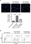
Migration of CD8+NKG2D+ T cells into alopecic skin. CD8+NKG2D T cells or CD8+NKG2D+ T cells were purified, CFSE-labeled and injected i.v. together with anti-CXCR3 mAb or control mAb. (A) Migration of CFSE-labeled T cells into AA skin grafts was evaluated by immunofluorescent microscopy. Data are the average of three mice, with at least three fields counted per mouse. (B) Representative FACS plots of single cell suspension of mouse skin. Shown are the percentages of the CFSE positive cells gated on CD45+CD3+CD8+. The relative frequency of CD8+CFSE positive cells was reduced by anti-CXCR3 mAb treatment. Data are representative of three independent experiments. ** indicates p<0.01, and *** indicates p<0.001.
FIGURE 5
FIGURE 5
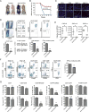
CXCR3 blockade prevents the onset of alopecia in AA skin grafted C3H/HeJ mice. Mice were treated beginning the day of grafting (n=5–10 mice per group). anti-CXCR3 mAb or isotype control IgG was administrated by i.p. injection (200 μg) two times weekly for 12 weeks. (A) The onset of alopecia was inhibited by administration of anti-CXCR3 mAb. (B) Time course of onset of AA in control mice and anti-CXCR3 treated mice was shown as weeks after grafting. (C) The expression of CD4, CD8, MHC class I and MHC class II in skin was significantly decreased in anti-CXCR3 treated mice compared to control mice. (D) Representative FACS plots of cell suspension of mouse skin. The frequencies of infiltrating CD45+ leukocytes and IFN-γ-producing CD8+NKG2D+ T cell in skin of anti-CXCR3 treated mice were significantly decreased compared to control mice. (E) CXCR3 ligand expression in skin from anti-CXCR3 treated mice and controls was analyzed using quantitative PCR. The expression of CXCR3 ligands in skin are significantly decreased in anti-CXCR3 treated mice compared to control mice. (F) Representative FACS plots of the SDLNs. Shown are the percentages of lymphocyte subsets. (G) The absolute numbers and the percentages of lymphocyte subsets in SDLNs are compared between the anti-CXCR3 treated group and control. Data are representative of two experiments with 10 mice total each group. Scale bar = 200μM. * indicates p<0.05, ** indicates p<0.01, and *** indicates p<0.001
FIGURE 6
FIGURE 6
CXCR3 blockade inhibits the proliferation of CD8+NKG2D+ T cells in SDLNs. C3H/HeJ mice skin grafted mice were treated as described in Methods. (A) Expression of CXCR3 ligands in SDLNs from anti-CXCR3 treated mice and controls was determined by quantitative RT-PCR. Data are indicated as the mean ± SEM of four mice in each group. (B) Frozen sections of cutaneous lymph nodes from each mouse in anti-CXCR3 treated or control groups were stained with anti-CXCL9 or anti-CXCL10 Abs. The expression of CXCR3 ligands in SDLNs were significantly decreased in anti-CXCR3 treated mice compared to control mice. Scale bar = 200μM. (C) The CD11c+MHC-II+ DCs in SDLNs were identified by gating on the T-cell-negative, B-cell-negative and NK-cell-negative population. The dot plots show that CD11c+MHC-II+ DCs within SDLNs strongly expressed CXCL9 and CXCL10. (D) Representative FACS plots showed the expression of CXCR3 was strongly associated with the expression of Ki-67 in either CD4+ or CD8+ T cell subsets within SDLNs of control skin grafted mice. Treatment with anti-CXCR3 reduced Ki67 expression in CD8 T cells. (E) CD8+ T cell proliferation was significantly inhibited by anti-CXCR3 treatment. CD8+ T cells purified from SDLN from C3H/HeJ mice with AA were CFSE stained and injected i.v. into C3H/HeJ AA mice recipients. Proliferation of the CFSE-stained CD8+ T cells within LNs (F) and spleen (G) was assessed by CFSE dilution. Data are representative of three independent experiments. * indicates p<0.05, ** indicates p<0.01, *** indicates p<0.001, n.s. indicates not significant.

Similar articles

Cited by

References

    1. Safavi KH, Muller SA, Suman VJ, Moshell AN, Melton LJ., 3rd Incidence of alopecia areata in Olmsted County, Minnesota, 1975 through 1989. Mayo Clin Proc. 1995;70:628–633. - PubMed
    1. Paus R, Nickoloff BJ, Ito T. A ‘hairy’ privilege. Trends Immunol. 2005;26:32–40. - PubMed
    1. Sun J, Silva KA, McElwee KJ, King LE, Jr, Sundberg JP. The C3H/HeJ mouse and DEBR rat models for alopecia areata: review of preclinical drug screening approaches and results. Exp Dermatol. 2008;17:793–805. - PMC - PubMed
    1. McElwee KJ, Gilhar A, Tobin DJ, Ramot Y, Sundberg JP, Nakamura M, Bertolini M, Inui S, Tokura Y, King LE, Jr, Duque-Estrada B, Tosti A, Keren A, Itami S, Shoenfeld Y, Zlotogorski A, Paus R. What causes alopecia areata? Exp Dermatol. 2013;22:609–626. - PMC - PubMed
    1. Xing L, Dai Z, Jabbari A, Cerise JE, Higgins CA, Gong W, de Jong A, Harel S, DeStefano GM, Rothman L, Singh P, Petukhova L, Mackay-Wiggan J, Christiano AM, Clynes R. Alopecia areata is driven by cytotoxic T lymphocytes and is reversed by JAK inhibition. Nat Med. 2014;20:1043–1049. - PMC - PubMed

Publication types

Substances