

Biophysical and Biochemical Characterization of Avian Secretory Component Provides Structural Insights into the Evolution of the Polymeric Ig Receptor
- PMID: 27412418
- PMCID: PMC4976031
- DOI: 10.4049/jimmunol.1600463
Biophysical and Biochemical Characterization of Avian Secretory Component Provides Structural Insights into the Evolution of the Polymeric Ig Receptor
Abstract
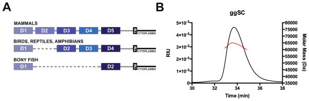
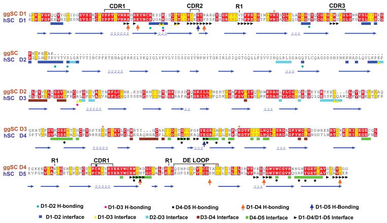
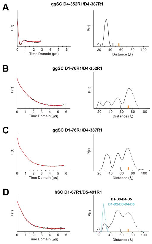
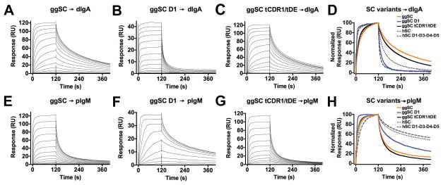
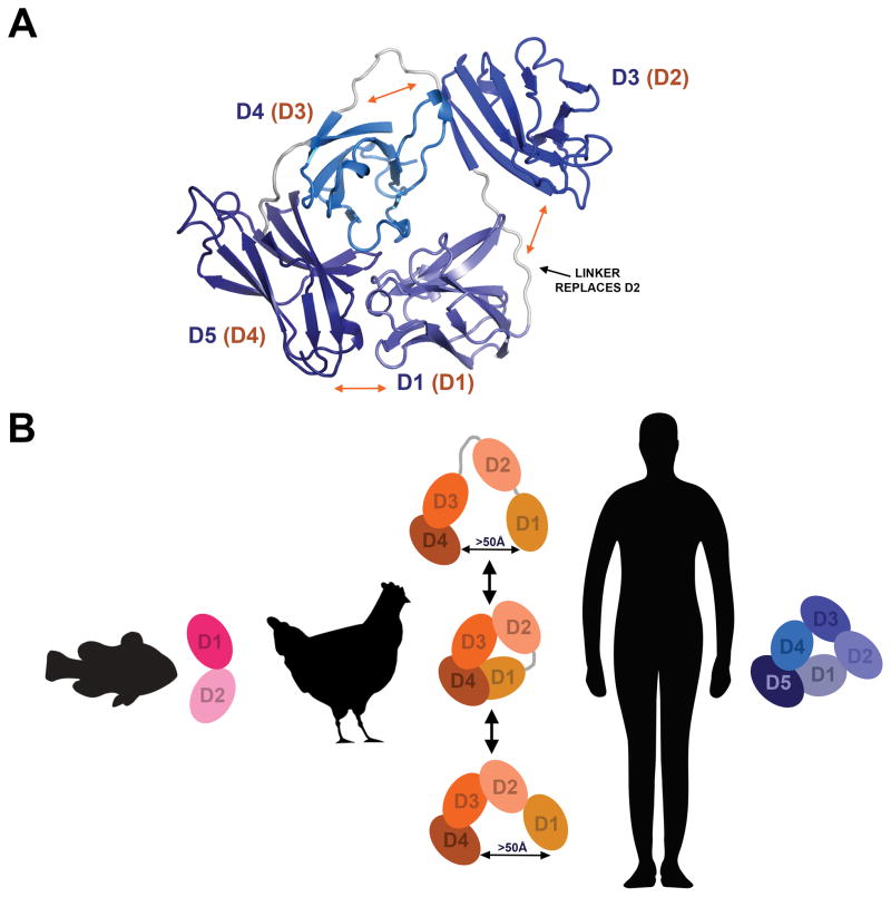
The polymeric Ig receptor (pIgR) transports polymeric Abs across epithelia to the mucosa, where proteolytic cleavage releases the ectodomain (secretory component [SC]) as an integral component of secretory Abs, or as an unliganded protein that can mediate interactions with bacteria. SC is conserved among vertebrates, but domain organization is variable: mammalian SC has five domains (D1-D5), whereas avian, amphibian, and reptilian SC lack the D2 domain, and fish SC lacks domains D2-D4. In this study, we used double electron-electron resonance spectroscopy and surface plasmon resonance binding studies to characterize the structure, dynamics, and ligand binding properties of avian SC, avian SC domain variants, and a human SC (hSC) variant lacking the D2 domain. These experiments demonstrated that, unlike hSC, which adopts a compact or "closed" domain arrangement, unliganded avian SC is flexible and exists in both closed and open states, suggesting that the mammalian SC D2 domain stabilizes the closed conformation observed for hSC D1-D5. Experiments also demonstrated that avian and mammalian pIgR share related, but distinct, mechanisms of ligand binding. Together, our data reveal differences in the molecular recognition mechanisms associated with evolutionary changes in the pIgR protein.
Copyright © 2016 by The American Association of Immunologists, Inc.
Figures
Similar articles
-
The structure and dynamics of secretory component and its interactions with polymeric immunoglobulins.Elife. 2016 Mar 4;5:e10640. doi: 10.7554/eLife.10640. Elife. 2016. PMID: 26943617 Free PMC article.
-
A functional polymeric immunoglobulin receptor in chicken (Gallus gallus) indicates ancient role of secretory IgA in mucosal immunity.Biochem J. 2004 Jun 15;380(Pt 3):669-76. doi: 10.1042/BJ20040200. Biochem J. 2004. PMID: 14992684 Free PMC article.
-
Mutational analysis of polymeric immunoglobulin receptor/ligand interactions. Evidence for the involvement of multiple complementarity determining region (CDR)-like loops in receptor domain I.J Biol Chem. 1994 Dec 16;269(50):31620-5. J Biol Chem. 1994. PMID: 7989333
-
Role of J chain in secretory immunoglobulin formation.Scand J Immunol. 2000 Sep;52(3):240-8. doi: 10.1046/j.1365-3083.2000.00790.x. Scand J Immunol. 2000. PMID: 10972899 Review.
-
Novel functions of the polymeric Ig receptor: well beyond transport of immunoglobulins.Trends Immunol. 2003 Feb;24(2):55-8. doi: 10.1016/s1471-4906(02)00031-5. Trends Immunol. 2003. PMID: 12547499 Review.
Cited by
-
Role of Polymeric Immunoglobulin Receptor in IgA and IgM Transcytosis.Int J Mol Sci. 2021 Feb 25;22(5):2284. doi: 10.3390/ijms22052284. Int J Mol Sci. 2021. PMID: 33668983 Free PMC article. Review.
-
The contribution of modern EPR to structural biology.Emerg Top Life Sci. 2018 Apr 20;2(1):9-18. doi: 10.1042/ETLS20170143. Emerg Top Life Sci. 2018. PMID: 33525779 Free PMC article.
-
Structural and Biochemical Requirements for Secretory Component Interactions with Dimeric IgA.J Immunol. 2024 Jul 15;213(2):226-234. doi: 10.4049/jimmunol.2300717. J Immunol. 2024. PMID: 38809110 Free PMC article.
-
Immunity in Sea Turtles: Review of a Host-Pathogen Arms Race Millions of Years in the Running.Animals (Basel). 2023 Feb 5;13(4):556. doi: 10.3390/ani13040556. Animals (Basel). 2023. PMID: 36830343 Free PMC article. Review.
-
The structures of secretory and dimeric immunoglobulin A.Elife. 2020 Oct 27;9:e56098. doi: 10.7554/eLife.56098. Elife. 2020. PMID: 33107820 Free PMC article.
References
-
- Kaetzel CS. Coevolution of Mucosal Immunoglobulins and the Polymeric Immunoglobulin Receptor: Evidence That the Commensal Microbiota Provided the Driving Force. ISRN Immunology. 2014;2014:20.
-
- Kaetzel CS. The polymeric immunoglobulin receptor: bridging innate and adaptive immune responses at mucosal surfaces. Immunol Rev. 2005;206:83–99. - PubMed
-
- Lindh E. Increased risistance of immunoglobulin A dimers to proteolytic degradation after binding of secretory component. J Immunol. 1975;114:284–286. - PubMed
Publication types
MeSH terms
Substances
Grants and funding
LinkOut - more resources
Full Text Sources
Other Literature Sources
Miscellaneous
