. 2016 Jul 26;113(30):E4397-406.
doi: 10.1073/pnas.1605578113. Epub 2016 Jul 13.

Endogenous N-acyl taurines regulate skin wound healing

Affiliations

Endogenous N-acyl taurines regulate skin wound healing

Oscar Sasso et al. Proc Natl Acad Sci U S A. .

Abstract

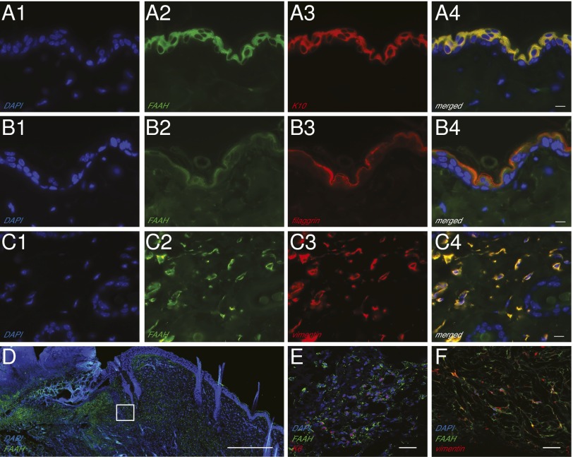
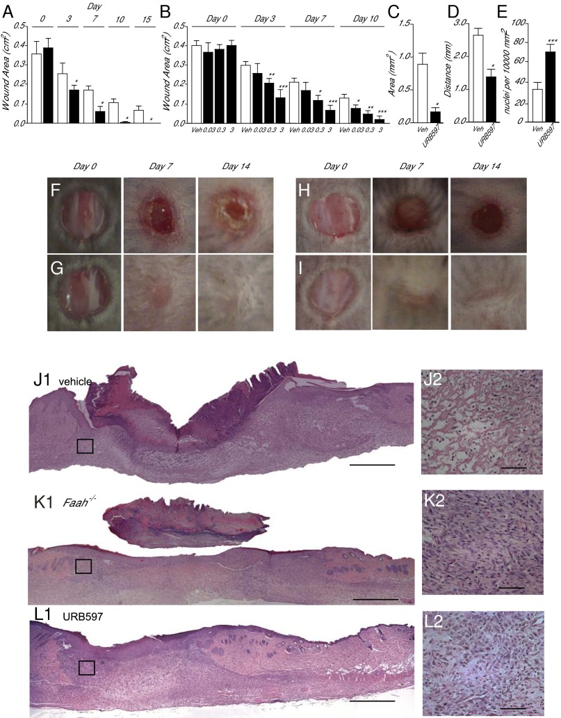
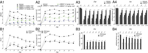
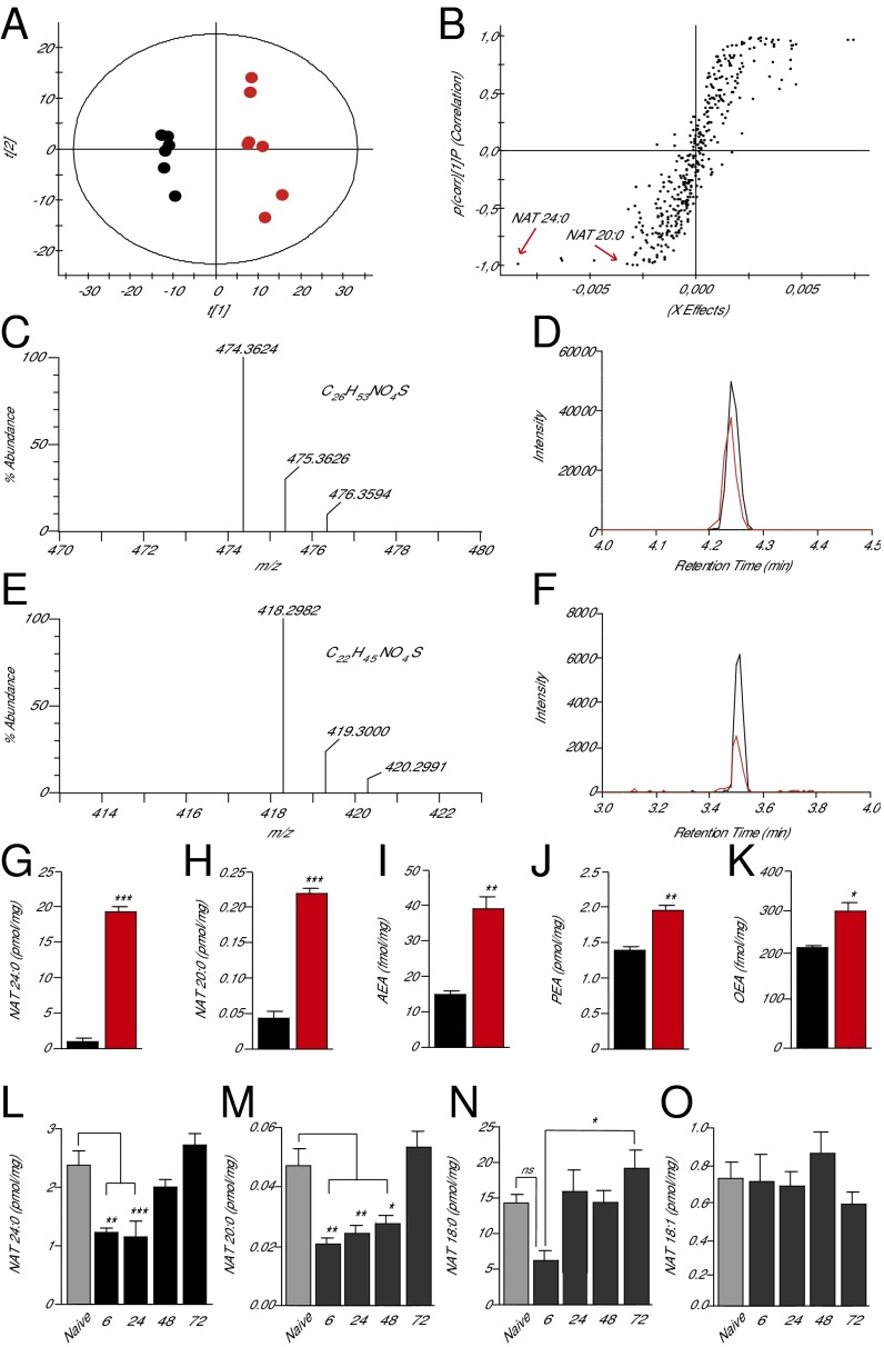
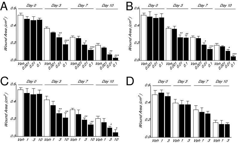
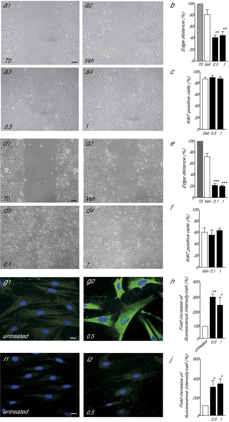
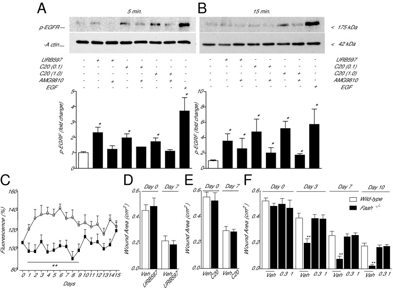
The intracellular serine amidase, fatty acid amide hydrolase (FAAH), degrades a heterogeneous family of lipid-derived bioactive molecules that include amides of long-chain fatty acids with taurine [N-acyl-taurines (NATs)]. The physiological functions of the NATs are unknown. Here we show that genetic or pharmacological disruption of FAAH activity accelerates skin wound healing in mice and stimulates motogenesis of human keratinocytes and differentiation of human fibroblasts in primary cultures. Using untargeted and targeted lipidomics strategies, we identify two long-chain saturated NATs-N-tetracosanoyl-taurine [NAT(24:0)] and N-eicosanoyl-taurine [NAT(20:0)]-as primary substrates for FAAH in mouse skin, and show that the levels of these substances sharply decrease at the margins of a freshly inflicted wound to increase again as healing begins. Additionally, we demonstrate that local administration of synthetic NATs accelerates wound closure in mice and stimulates repair-associated responses in primary cultures of human keratinocytes and fibroblasts, through a mechanism that involves tyrosine phosphorylation of the epidermal growth factor receptor and an increase in intracellular calcium levels, under the permissive control of transient receptor potential vanilloid-1 receptors. The results point to FAAH-regulated NAT signaling as an unprecedented lipid-based mechanism of wound-healing control in mammalian skin, which might be targeted for chronic wound therapy.

Keywords: FAAH; FAEs; N-acyl taurines; fibroblasts; keratinocytes.


Conflict of interest statement

Conflict of interest statement: M.M. and D.P. are inventors on issued patents, assigned to the University of California and the Istituto Italiano di Tecnologia, for URB597 and other fatty acid amide hydrolase inhibitors.

Figures

Fig. 1.
Fig. 1.
Localization of FAAH in adult mouse skin. (AC) Representative double immunofluorescence images for FAAH, the keratinocyte markers cytokeratin-10 (K10) (A1A4) and filaggrin (B1B4), and the fibroblast marker vimentin (C1C4) in intact skin. (Scale bars, 10 µm.) (D) Immunofluorescence localization of FAAH in wounded skin. The highlighted square is magnified in E and F, to show the presence of immunoreactive FAAH in keratinocytes (cytokeratin-5; E) and fibroblasts (vimentin; F). [Scale bars, 500 µm (D) and 20 µm (E and F)]. FAAH is shown in green, and all other markers in red; colocalization (A4, B4, and C4) is shown in yellow; nuclei are stained with DAPI (blue). An antibody selective for FAAH-1, the only FAAH isoform present in rodent tissues, was used in these experiments.
Fig. 2.
Fig. 2.
FAAH regulates healing of excisional skin wounds in mice. (A) Time course (days) of wound healing in wild-type mice (open bars) and Faah−/− mice (filled bars). (B) Time course of wound healing in wild-type mice treated with vehicle (open bars) or with various topical doses (%; wt/vol) of the FAAH inhibitor URB597 (filled bars). (CE) Morphometric analyses showing the effects of vehicle (open bars) or URB597 (3%, wt/vol) (filled bars) on wound area (C), distance between migration tongues (D), and cell density in the dermis (E). (FI) Representative images illustrating skin wound healing in drug-naïve wild-type mice (F), drug-naïve Faah−/− mice (G), and wild-type mice treated either with vehicle (H) or URB597 (3%, wt/vol; I). (JL) Hematoxylin/eosin staining of skin sections from drug-naïve wild-type mice (J1), drug-naïve Faah−/− mice (K1), and wild-type mice treated with URB597 (3%, wt/vol; L1). The highlighted squares are magnified in J2, K2, and L2. [Scale bars, 500 µm (J1, K1, and L1) and 50 µm (J2, K2, and L2).] Skin sections were prepared 7 d after wounding. Data are expressed as mean ± SEM (n = 9). *P < 0.05; **P < 0.01; ***P < 0.001 (compared with vehicle or wild-type mice, two-tailed Student’s t test).
Fig. 3.
Fig. 3.
FAAH regulates recovery of paw tissue structure and function after a surgical incision. (A) Time course (days) of recovery in mice treated with the FAAH inhibitor URB597 (1, 3, and 10 mg/kg, oral). The following parameters were evaluated: wound closure and scar formation (A1), paw edema (A2), heat hyperalgesia (A3), and mechanical allodynia (A4). (B) Time course of recovery in wild-type and Faah−/− mice: wound closure (B1), edema (B2), hyperalgesia (B3), and allodynia (B4). Data are expressed as mean ± SEM (n = 9). *P < 0.05; **P < 0.01; ***P < 0.001 (compared with vehicle or wild-type mice, two-way ANOVA).
Fig. 4.
Fig. 4.
Identification of preferred FAAH substrates in mouse skin. (A) Score plot from PCA of skin lipidomics profiles of wild-type mice (black circles) and Faah−/− mice (red circles) acquired in negative-ion mode; t[1] and t[2], principal component 1 and 2, respectively. (B) Sigmoidal plot of OPLS-DA analysis of the negative-ion dataset. Signals strongly contributing to class separation are those found at the bottom left (Faah−/− mice) or top right (wild-type mice) of the plot. Details for each signal are reported in SI Appendix, Table S1. (CF) High-resolution mass spectra, calculated molecular formulae, and LC/MS tracings for native and synthetic NAT(24:0) (C and D) and NAT(20:0) (E and F). Native compounds are shown in red, authentic standards are shown in black. (GK) Quantification of NAT(24:0) (G), NAT(20:0) (H), anandamide (AEA) (I), PEA (J), and OEA (K) in skin of wild-type (black bars) and Faah−/− (red bars) mice. (LO) Time course of changes in the levels of NAT(24:0) (L), NAT(20:0) (M), NAT(18:0) (N), and NAT(18:1) (O) in skin tissue surrounding excision wounds (solid bars) or from unwounded mice (shaded bars). Data are expressed as mean ± SEM (n = 3). *P < 0.05; **P < 0.01; ***P < 0.001 (compared with wild-type mice, two-tailed Student’s t test; n = 5); #P < 0.05 (one-way ANOVA); ns, not significant.
Fig. 5.
Fig. 5.
Synthetic NATs accelerate skin healing in mice. (A and B) Time course (days) of wound closure in wild-type mice treated daily with topical applications of vehicle (open bars) or synthetic NAT(24:0) (A), NAT(20:0) (B), PEA (C), or anandamide (D) (%; filled bars). Data are expressed as mean ± SEM (n = 9). *P < 0.05; **P < 0.01; ***P < 0.001 (compared with vehicle or wild-type mice, two-tailed Student’s t test).
Fig. 6.
Fig. 6.
FAAH controls human keratinocyte migration and human fibroblasts transdifferentiation into myofibroblasts. (A and B) Representative image illustrating the motogenic effects of the FAAH inhibitor URB597 (0.5 or 1 μM; 18 h) in primary cultures of human keratinocytes (A) and quantitative analysis of leading edges distance (B). Data are expressed as percent of vehicle-treated control cultures (mean ± SEM). (C) Effects of URB597 (48 h) on keratinocyte proliferation. (DF) Effects of synthetic NAT(20:0) on keratinocyte migration (18 h) (D and E) and keratinocyte proliferation (48 h) (F). (GJ) Effects of URB597 (0.5 μM, 48 h) on the expression of α-smooth muscle actin (G and H) and procollagen type I (I and J) in human fibroblasts. Data are expressed as mean ± SEM (n = 3). (Scale bars, 20 µm.) *P < 0.05; **P < 0.01; ***P < 0.001 (compared with vehicle mice, two-tailed Student’s t test).
Fig. 7.
Fig. 7.
Signal transduction events initiated by FAAH-regulated NAT signaling in human keratinocytes. (A and B) Western blot analyses of primary cultures of human keratinocytes 5 min (A) and 15 min (B) after addition of the FAAH inhibitor URB597 or synthetic NAT(20:0) (C20 0.1 and 1 μM), in the absence or presence of the TRPV-1 antagonist AMG9810 (5 μM). (A, Upper, and B, Upper) Representative blots. (A, Lower, and B, Lower) Quantification of data from three independent experiments. EGF was used as positive control and β-actin as loading control. (C) Intracellular calcium levels (fluo-3 fluorescence) in human keratinocytes incubated with URB597 (1 μM; open circles) or URB597 plus TRPV-1 antagonist AMG9810 (5 μM; filled squares). Vehicle and AMG9810 had no effect on calcium levels when applied alone. (D) Effects of vehicle (open bars) or URB597 [3% (wt/vol); filled bars] on wound healing in Trpv1−/− mice. (E) Effects of vehicle (open bars) or NAT(20:0) [0.01% (wt/vol); filled bars] on wound healing in Trpv1−/− mice. In D and E, wound areas were measured 0 and 7 d after wounding. (F) Time-course of wound healing in wild-type mice treated with vehicle (open bars) and Faah−/− mice (filled bars) treated with vehicle or AMG9810 [0.3 or 1% (wt/vol)]. Data are expressed as mean± SEM (n = 3 for keratinocyte experiments, and n = 9 for in vivo experiments). *P < 0.05; **P < 0.01; ***P < 0.001 (compared with controls).

References

    1. Bíró T, Tóth BI, Haskó G, Paus R, Pacher P. The endocannabinoid system of the skin in health and disease: Novel perspectives and therapeutic opportunities. Trends Pharmacol Sci. 2009;30(8):411–420. - PMC - PubMed
    1. Kendall AC, Nicolaou A. Bioactive lipid mediators in skin inflammation and immunity. Prog Lipid Res. 2013;52(1):141–164. - PubMed
    1. Blankman JL, Cravatt BF. Chemical probes of endocannabinoid metabolism. Pharmacol Rev. 2013;65(2):849–871. - PMC - PubMed
    1. Devane WA, et al. Isolation and structure of a brain constituent that binds to the cannabinoid receptor. Science. 1992;258(5090):1946–1949. - PubMed
    1. Piomelli D, Sasso O. Peripheral gating of pain signals by endogenous lipid mediators. Nat Neurosci. 2014;17(2):164–174. - PMC - PubMed

Publication types

MeSH terms