. 2016 Oct 6;128(14):1789-1799.
doi: 10.1182/blood-2016-04-709634. Epub 2016 Jul 13.

The antigenic complex in HIT binds to B cells via complement and complement receptor 2 (CD21)

Affiliations

The antigenic complex in HIT binds to B cells via complement and complement receptor 2 (CD21)

Sanjay Khandelwal et al. Blood. .

Abstract

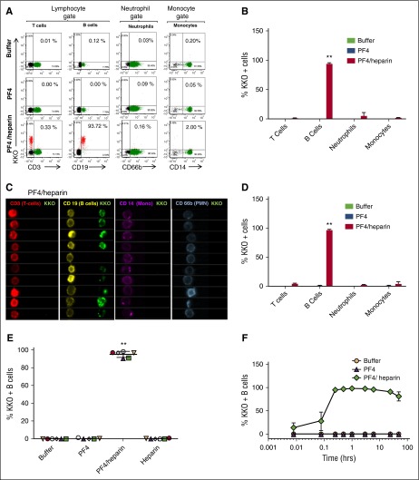
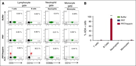
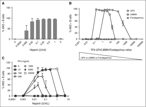
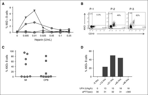
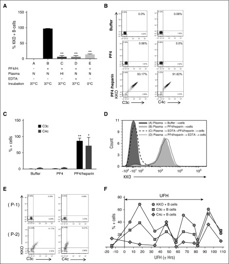
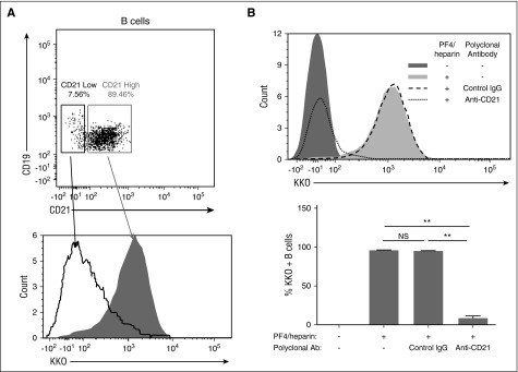
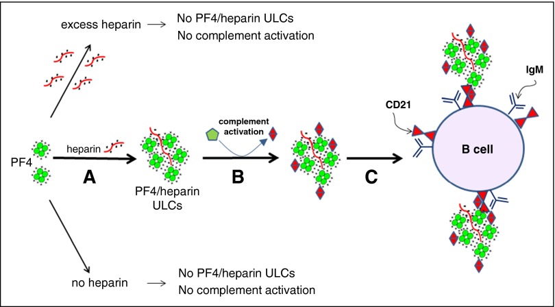
Heparin-induced thrombocytopenia is a prothrombotic disorder caused by antibodies to platelet factor 4 (PF4)/heparin complexes. The mechanism that incites such prevalent anti-PF4/heparin antibody production in more than 50% of patients exposed to heparin in some clinical settings is poorly understood. To investigate early events associated with antigen exposure, we first examined the interaction of PF4/heparin complexes with cells circulating in whole blood. In healthy donors, PF4/heparin complexes bind preferentially to B cells (>90% of B cells bind to PF4/heparin in vitro) relative to neutrophils, monocytes, or T cells. Binding of PF4 to B cells is heparin dependent, and PF4/heparin complexes are found on circulating B cells from some, but not all, patients receiving heparin. Given the high proportion of B cells that bind PF4/heparin, we investigated complement as a mechanism for noncognate antigen recognition. Complement is activated by PF4/heparin complexes, co-localizes with antigen on B cells from healthy donors, and is present on antigen-positive B cells in patients receiving heparin. Binding of PF4/heparin complexes to B cells is mediated through the interaction between complement and complement receptor 2 (CR2 [CD21]). To the best of our knowledge, these are the first studies to demonstrate complement activation by PF4/heparin complexes, opsonization of PF4/heparin to B cells via CD21, and the presence of complement activation fragments on circulating B cells in some patients receiving heparin. Given the critical contribution of complement to humoral immunity, our observations provide new mechanistic insights into the immunogenicity of PF4/heparin complexes.


Figures

Figure 1
Figure 1
PF4/heparin complexes bind preferentially to B cells in the peripheral blood. Whole blood from healthy donors was incubated with buffer or antigen (PF4 with or without heparin) followed by staining for cell-specific markers and KKO. Mean ± standard deviation (SD) of 3 independent experiments is shown. (A) Flow cytometric analyses of peripheral blood leukocytes. Leukocyte subpopulations were defined by forward scatter and side scatter characteristics (lymphocytes, neutrophils, and monocytes). Gated subpopulations were identified by cell-surface markers for T lymphocytes (CD3), B lymphocytes (CD19), neutrophils (CD66b), and monocytes (CD14) on the x-axis and for KKO staining on the y-axis for each antigen. The percentage of KKO-positive cells for each cell lineage appears in the upper right corner of each graph. (B) Graph of cell lineage–specific staining of flow data from (A). Binding of KKO to cell lineages incubated with antigen is shown. (C) ImageStream analysis of PF4/heparin binding to B cells. Cell-surface binding of PF4/heparin complexes is shown by imaging flow cytometry using fluorescently labeled cell-surface markers (CD3-APC, CD19-PE, CD14-APC, and CD66b-APC) and KKO-AF488 (green). For clarity, colors were reassigned to show T cells in red, B cells in yellow, monocytes in purple, and neutrophils in blue. (D) Graph of cell lineage–specific staining of ImageStream data from (C). (E) PF4/heparin complexes bind preferentially to B cells from multiple healthy donors. CD19+ cells were gated for KKO binding for various antigens. The graphs show percentage of KKO-positive B cells (mean ± SD) in various conditions from 6 healthy donors. Each colored symbol represents % KKO + B cells in an individual donor. (F) Kinetics of PF4/heparin binding to B cells. Whole blood was incubated with antigen for defined time points and stained with KKO/CD19. For each condition, time is shown on the x-axis and binding of KKO shown on the y-axis. **P < .005 compared with other data in graph.
Figure 2
Figure 2
PRT/heparin complexes show similar selective binding to B cells. Whole blood from a representative healthy donor was incubated with buffer or antigen (PRT with or without heparin) followed by staining for cell-specific markers and ADA, a monoclonal antibody to PRT/heparin complexes. Mean ± SD of 3 independent experiments is shown. (A) Flow cytometric analyses of peripheral blood leukocytes incubated with buffer, PRT, or PRT/heparin. Flow cytometry was performed on whole blood. Leukocyte subpopulations were defined by forward and side scatter characteristics (lymphocytes, neutrophils, and monocytes). Gated subpopulations were identified by cell-surface markers for T lymphocytes (CD3), B lymphocytes (CD19), neutrophils (CD66b), and monocytes (CD14) on the x-axis and for ADA staining on the y-axis for each antigen. The percentage of ADA-positive cells for each cell lineage appears in the upper right corner of each graph. (B) Graph of cell lineage–specific staining of flow data shown in (A). Binding of ADA to cell lineages incubated with antigen is shown. **P < .005 compared with other data in graph.
Figure 3
Figure 3
PF4/heparin complexes bind B cells in a heparin concentration–dependent manner. Whole blood was incubated with various concentrations of PF4 in combination with UFH, LMWH, or fondaparinux as indicated. Cells were stained with CD19 and KKO and the percentage of B cells binding with KKO under each condition is shown on the y-axis. (A) PF4/heparin binding to B cells is heparin dependent. Whole blood was incubated with a fixed amount of PF4 (25 µg/mL) and various concentrations of UFH (0-5 U/mL). Shown are the mean ± SD for percentage of KKO-positive B cells as a function of UFH concentration from 4 independent experiments. (B) B cell binding of complexes formed with PF4 and UFH, LMWH, or fondaparinux. Whole blood was incubated with a fixed amount of PF4 (25 µg/mL) and various concentrations of UFH, LMWH, or fondaparinux. Shown are the mean ± SD for percentage of KKO-positive B cells as a function of PF4 and UFH, LMWH, and fondaparinux molar ratios from 3 independent experiments. (C) B cell binding of PF4/heparin ULCs increases with PF4/heparin concentrations. Whole blood was incubated with increasing amounts of PF4 (0-25 000 ng/mL) and increasing UFH concentrations (0-5 U/mL). KKO binding for a given PF4 concentration is shown as a function of heparin concentration. Shown are the mean ± SD for percentage of KKO-positive B cells from 3 independent experiments.
Figure 4
Figure 4
B cells bind PF4/heparin complexes ex vivo and in vivo. (A) Heparin displaces variable amounts of PF4 leading to B-cell binding of PF4/heparin complexes in some healthy subjects. Blood from 6 healthy donors was incubated with various concentrations of UFH as indicated. Binding of PF4/heparin complexes to B cells as assessed by binding of KKO is shown. (B) Detection of PF4/heparin complex bound circulating B cells in 3 patients receiving heparin. Blood was stained for CD19 and KKO. Dot plots show variation in binding of KKO to lymphocyte-gated populations. (C) Variations in KKO binding of PF4/heparin complexes to B cells in vivo in patients receiving UFH for medical indications (MI; n = 8) or for cardiopulmonary bypass surgery (CPB; n = 8). Each symbol represents B cell binding in an individual patient. The y-axis indicates maximal percentage of KKO-bound B cells. (D) In vivo B-cell binding is heparin dependent. A patient undergoing UFH therapy for treatment of cerebral venous thrombosis was monitored for percentage of KKO-positive B cells over 48 hours of UFH therapy. Time from start of heparin therapy, UFH dose, and activated partial thromboplastin time (aPTT) are shown.
Figure 5
Figure 5
Complement mediates binding of PF4/heparin complexes to B cells. (A) Effect of complement inhibition on PF4/heparin binding to B cells. Plasma and blood cells were separated from blood by centrifugation, subjected to conditions associated with complement inactivation (heat inactivation and treatment with 10 mM EDTA or ice), and exposed to antigen or buffer. Conditions A to E correspond to various incubation conditions. Mean ± SD from 3 independent experiments. **P < .005 compared with condition B. N, normal; HI, heat inactivated. (B) and (C) PF4/heparin binding to B cells correlates with C3c/C4c deposition. Percentage of double-positive cells, which represent KKO and C3c or C4c binding, appears in the right upper quadrant of dot plots (B) and as quantified in (C) from 3 independent experiments. *P < .05, and **P < .005 compared with buffer condition. (D) Complement fixation occurs primarily in the solution phase and not on the B-cell surface. Plasma and blood cells were separated, and sequence of B-cell binding was determined as described in “Methods.” Overlay histogram for KKO staining on the CD19-gated B cells is shown by the sequence of incubations as indicated in the key. (E) Binding of PF4/heparin complexes and complement to B cells from heparinized patients. Blood from 2 patients (P-1 and P-2) was stained concurrently for CD19, KKO, C3c, or C4c. Dot plots show binding of KKO and C3c/C4c binding on CD19+ gated cells. (F) Binding of PF4/heparin, C3c, and C4c to B cells over time in a patient treated with heparin. Shown is the percentage of KKO-positive/C3c-positive/C4c-positive B cells in the circulation of a heparinized patient during the course of heparin therapy. The x-axis shows time from start of heparin therapy, the y-axis shows % KKO + B cells, and the 2-headed arrow in the figure indicates duration of UFH therapy.
Figure 6
Figure 6
Complement-containing PF4/heparin complexes bind to CR2 (CD21) on B cells. (A) Binding of PF4/heparin to peripheral blood B cells correlates with CD21 expression. Whole blood was incubated with PF4/heparin and stained with labeled antibodies to CD19, CD21, and KKO. Top panel: gating of B cells based on CD21 expression; bottom panel: overlay histogram of KKO staining as a function of CD21 expression as low- (open curve with solid line) or high- (shaded area) expressing B cells. (B) CD21 mediates binding of complement-coated PF4/heparin complexes to B cells. Blood was incubated with anti-CD21 or control IgG before the addition of PF4/heparin. Top panel: a representative overlay histogram is shown for KKO staining of B cells with anti-CD21 or control IgG. Bottom panel: summary of results (mean ± SD) from 3 experiments for PF4/heparin binding to B cells after expression of CD21 was blocked with a polyclonal anti-CD21 antibody. **P < .005.
Figure 7
Figure 7
Activation of complement by PF4/heparin complexes and binding to B-cell CD21. (A) PF4 and heparin interact over a narrow range of molar ratios to generate ULCs. PF4 alone or PF4 with excess of heparin do not make ULCs. (B) ULCs activate complement and bind complement activation products (C3/C4). (C) Complement-coated ULCs bind to a B cell via CR2 (CD21).

Comment in

Similar articles

Cited by

References

    1. Suvarna S, Espinasse B, Qi R, et al. Determinants of PF4/heparin immunogenicity. Blood. 2007;110(13):4253–4260. - PMC - PubMed
    1. Degn SE, Thiel S. Humoral pattern recognition and the complement system. Scand J Immunol. 2013;78(2):181–193. - PubMed
    1. Suvarna S, Qi R, Hollingsworth JW, Arepally GM. Platelet factor 4-heparin complexes trigger immune responses independently of the MyD88 pathway. Br J Haematol. 2008;142(4):671–673. - PubMed
    1. Prechel MM, Walenga JM. Complexes of platelet factor 4 and heparin activate Toll-like receptor 4. J Thromb Haemost. 2015;13(4):665–670. - PubMed
    1. Carroll MC. The complement system in regulation of adaptive immunity. Nat Immunol. 2004;5(10):981–986. - PubMed

Publication types

MeSH terms