. 2016 Jul 13;5(7):e003621.
doi: 10.1161/JAHA.116.003621.

Obesity Is Associated With Progression of Atherosclerosis During Statin Treatment

Affiliations

Obesity Is Associated With Progression of Atherosclerosis During Statin Treatment

Veit Sandfort et al. J Am Heart Assoc. .

Abstract

Background: This study aimed to determine the relationship of statin therapy and cardiovascular risk factors to changes in atherosclerosis in the carotid artery.

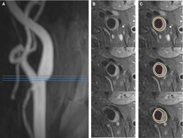
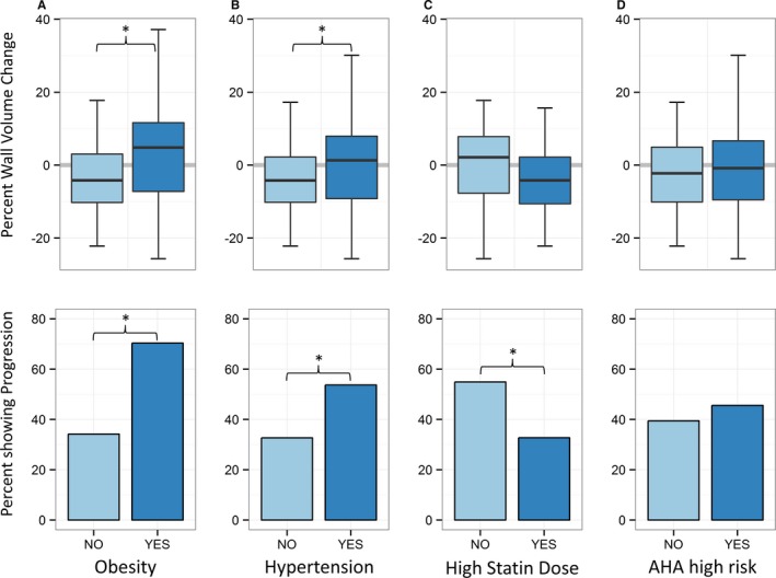
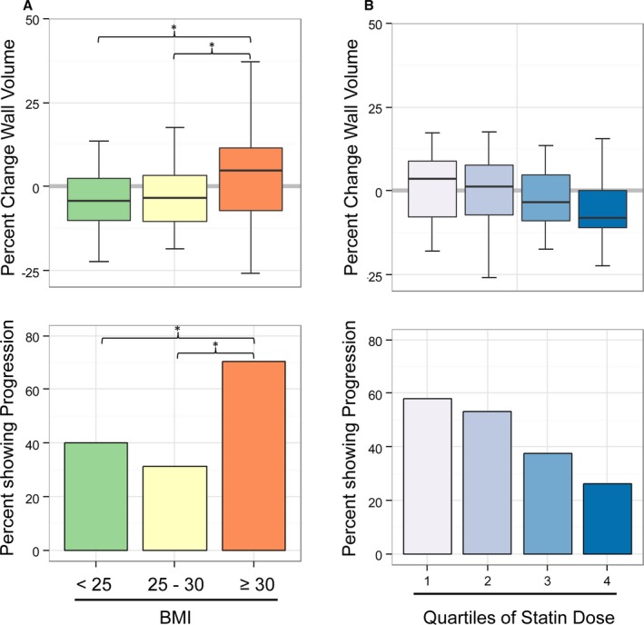
Methods and results: Carotid magnetic resonance imaging was used to evaluate 106 hyperlipidemic participants at baseline and after 12 months of 3-hydroxy-3-methylglutaryl coenzyme A reductase inhibitor (statin) treatment. Multivariable logistic regression was used to determine factors associated with progression (change in carotid wall volume >0) or regression (change ≤0) of carotid atherosclerosis. Computed tomography coronary calcium scores were obtained at baseline for all participants. The median age was 65 years (interquartile range 60-69 years), and 63% of the participants were male. Body mass index >30, elevated C-reactive protein, and hypertension were associated with increased carotid wall volume (obesity: odds ratio for progression 4.6, 95% CI 1.8-12.4, P<0.01; C-reactive protein: odds ratio for progression 2.56, 95% CI 1.17-5.73, P=0.02; hypertension: odds ratio 2.4, 95% CI 1.1-5.3, P<0.05). Higher statin dose was associated with regression of carotid wall volume (P<0.05). In multivariable analysis, obesity remained associated with progression (P<0.01), whereas statin use remained associated with regression (P<0.05). Change in atheroma volume in obese participants was +4.8% versus -4.2% in nonobese participants (P<0.05) despite greater low-density lipoprotein cholesterol reduction in obese participants.

Conclusions: In a population with hyperlipidemia, obese patients showed atheroma progression despite optimized statin therapy.

Clinical trial registration: URL: http://www.clinicaltrials.gov. Unique identifier: NCT01212900.

Keywords: carotid artery; carotid magnetic resonance imaging; obesity.


Figures

Figure 1
Figure 1
Assessment of carotid wall volume. A, Magnetic resonance angiography shows mild wall irregularity of the internal carotid artery. B, Representative axial slices show a region of wall thickening and plaque formation. C, The vessel boundaries were traced in multiple slices, and the volume was calculated. Three continuous axial slices are shown; 5 slices were acquired and analyzed to obtain the wall volume.
Figure 2
Figure 2
LDL cholesterol values at baseline and at 6 and 12 months. Statin‐naïve baseline values were calculated using the baseline LDL, statin dose, and expected LDL‐lowering effects.21 A, During the treatment protocol, a further decrease in LDL values occurred (*P<0.01). B, Of note, the LDL reduction was more pronounced in obese participants (yellow) compared with nonobese participants (green). BL indicates baseline; LDL, low‐density lipoprotein.
Figure 3
Figure 3
Upper row shows univariate comparisons of continuous carotid wall volume change. A, Change for participants with low BMI (light blue) vs high BMI (dark blue). B, Change in participants with (dark blue) and without (light blue) hypertension. C, Change in participants with a high statin dose (≥40 mg Simvastatin, dark blue) and low statin dose (light blue). D, Change in participants with an AHA risk <7.5% (light blue) or ≥7.5% (dark blue). Lower row shows corresponding percentages of participants experiencing progression of carotid wall volume (change >0). Obese and hypertensive participants had greater wall volume change and more frequently showed carotid artery disease progression (*P<0.05). AHA indicates American Heart Association; BMI, body mass index.
Figure 4
Figure 4
A, Change in carotid wall volume in normal‐weight, overweight, and obese participants. Obese patients showed wall volume increase (progression) opposed to normal‐weight and overweight participants (*P<0.05). B, Change in carotid wall volume in relation to quartiles of statin dose. There was a stepwise reduction in wall volume change with increasing dose (inverse correlation, P=0.038). BMI indicates body mass index.

References

    1. Global, regional, and national age‐sex specific all‐cause and cause‐specific mortality for 240 causes of death, 1990–2013: a systematic analysis for the Global Burden of Disease Study 2013. Lancet. 2015;385:117–171. - PMC - PubMed
    1. Kajinami K, Akao H, Polisecki E, Schaefer EJ. Pharmacogenomics of statin responsiveness. Am J Cardiol. 2005;96:65K–70K; discussion 34K‐35K. - PubMed
    1. Kataoka Y, St John J, Wolski K, Uno K, Puri R, Tuzcu EM, Nissen SE, Nicholls SJ. Atheroma progression in hyporesponders to statin therapy. Arterioscler Thromb Vasc Biol. 2015;35:990–995. - PubMed
    1. Martin SS, Sperling LS, Blaha MJ, Wilson PWF, Gluckman TJ, Blumenthal RS, Stone NJ. Clinician‐patient risk discussion for atherosclerotic cardiovascular disease prevention importance to implementation of the 2013 ACC/AHA Guidelines. J Am Coll Cardiol. 2015;65:1361–1368. - PMC - PubMed
    1. Zavodni AE, Wasserman BA, McClelland RL, Gomes AS, Folsom AR, Polak JF, Lima JA, Bluemke DA. Carotid artery plaque morphology and composition in relation to incident cardiovascular events: the Multi‐Ethnic Study of Atherosclerosis (MESA). Radiology. 2014;271:381–389. - PMC - PubMed

Publication types

MeSH terms

Substances

Associated data