. 2016 Jul 13;36(28):7428-40.
doi: 10.1523/JNEUROSCI.1114-16.2016.

Microbiota Dysbiosis Controls the Neuroinflammatory Response after Stroke

Affiliations

Microbiota Dysbiosis Controls the Neuroinflammatory Response after Stroke

Vikramjeet Singh et al. J Neurosci. .

Abstract

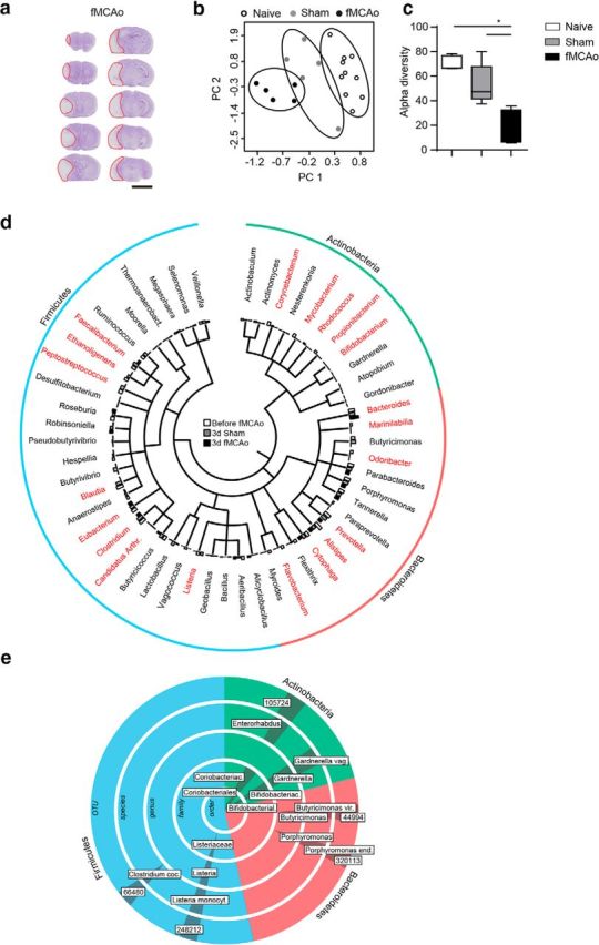
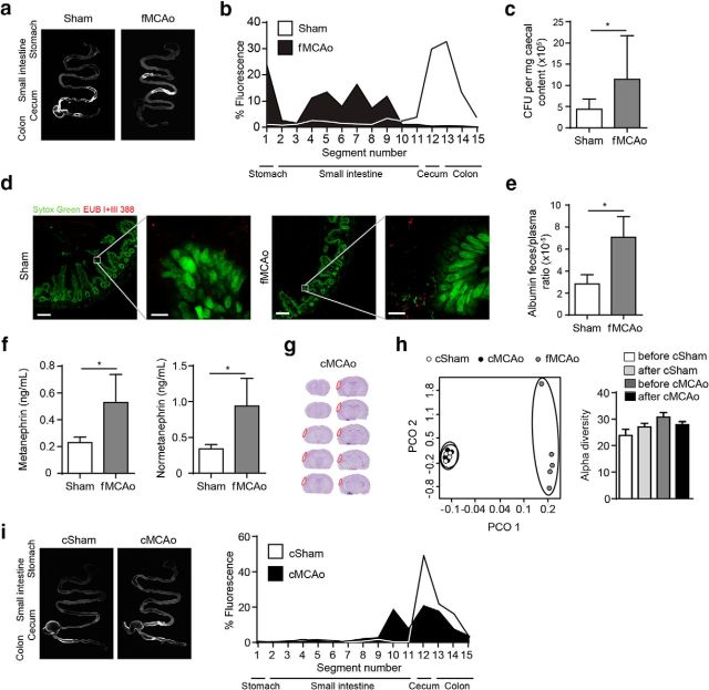
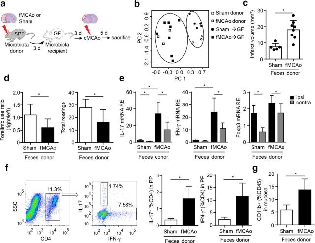
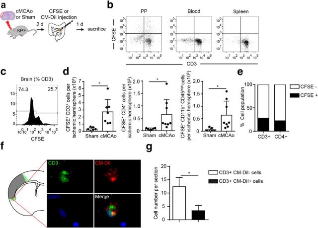
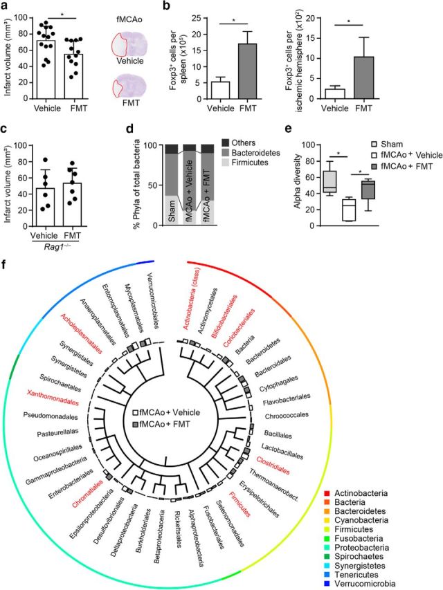
Acute brain ischemia induces a local neuroinflammatory reaction and alters peripheral immune homeostasis at the same time. Recent evidence has suggested a key role of the gut microbiota in autoimmune diseases by modulating immune homeostasis. Therefore, we investigated the mechanistic link among acute brain ischemia, microbiota alterations, and the immune response after brain injury. Using two distinct models of acute middle cerebral artery occlusion, we show by next-generation sequencing that large stroke lesions cause gut microbiota dysbiosis, which in turn affects stroke outcome via immune-mediated mechanisms. Reduced species diversity and bacterial overgrowth of bacteroidetes were identified as hallmarks of poststroke dysbiosis, which was associated with intestinal barrier dysfunction and reduced intestinal motility as determined by in vivo intestinal bolus tracking. Recolonizing germ-free mice with dysbiotic poststroke microbiota exacerbates lesion volume and functional deficits after experimental stroke compared with the recolonization with a normal control microbiota. In addition, recolonization of mice with a dysbiotic microbiome induces a proinflammatory T-cell polarization in the intestinal immune compartment and in the ischemic brain. Using in vivo cell-tracking studies, we demonstrate the migration of intestinal lymphocytes to the ischemic brain. Therapeutic transplantation of fecal microbiota normalizes brain lesion-induced dysbiosis and improves stroke outcome. These results support a novel mechanism in which the gut microbiome is a target of stroke-induced systemic alterations and an effector with substantial impact on stroke outcome.

Significance statement: We have identified a bidirectional communication along the brain-gut microbiota-immune axis and show that the gut microbiota is a central regulator of immune homeostasis. Acute brain lesions induced dysbiosis of the microbiome and, in turn, changes in the gut microbiota affected neuroinflammatory and functional outcome after brain injury. The microbiota impact on immunity and stroke outcome was transmissible by microbiota transplantation. Our findings support an emerging concept in which the gut microbiota is a key regulator in priming the neuroinflammatory response to brain injury. These findings highlight the key role of microbiota as a potential therapeutic target to protect brain function after injury.

Keywords: T cells; inflammation; microbiota; stroke.


Figures

Figure 1.
Figure 1.
Severe stroke induces microbiota dysbiosis. a, Representative images of cresyl-violet-stained coronal brain sections 3 d after fMCAo. In each section, the infarct area is outlined in red 5 mm). b, Principal component (PC) analysis of the intestinal microbiota by taxonomic abundance patterns in naive mice (before sham or fMCAo surgery) and after sham and fMCAo surgery. c, Quantitative analysis of α diversity confirms significantly reduced species diversity of the gut microbiota after brain injury in the fMCAo model (Mann–Whitney U test). d, Phylogenetic tree illustrating the distribution of the identified bacterial genera within the most abundant phyla (Actinobacteria, Bacteroidetes, and Firmicutes) comparing post-fMCAo mice with sham-operated and before-fMCAo mice. Genera that were significantly altered in the fMCAo group compared with the sham and before-fMCAo groups are indicated in red (ANOVA, n = 5 mice per group). e, LEfSe algorithm analysis was performed for indicator taxa analysis identifying features that are statistically different between sham- and fMCAo-operated mice. Labels in boxes define significantly regulated operational taxonomic units (OTU, numbers) and higher order taxa, respectively.
Figure 2.
Figure 2.
Lesion severity determines gastrointestinal dysfunction. a, Intestinal motility was measured 24 h after fMCAo or sham surgery. Representative fluorescence images of the complete gastrointestinal tract 60 min after gastric instillation of FITC-dextran showing retention of the fluorescent bolus in the upper gastrointestinal tract as a marker of severely impaired motility after fMCAo compared with sham mice. Arrowheads indicate cecum. b, Quantification of fluorescence intensity in intestinal segments. Note the retention of fluorescence signal in the upper gastrointestinal tract after fMCAo (n = 8 per group, 3 individual experiments). c, Number of colony forming units (CFUs) per milligram of murine cecal content cultured under anaerobic conditions after sham or fMCAo surgery (n = 8–9 mice per group, 2 independent experiments). d, No bacterial invasion into lamina propria was found in transverse sections of mouse ileum, which were hybridized using a general bacteria-specific EUB338 probe with Sytox green counterstaining. Left panels: Magnification, 20×; scale bars, 100 μm; right panels: magnification, 63×, scale bars, 10 μm. e, Albumin concentrations were determined by ELISA and are represented as the ratio of the concentrations in feces and plasma from sham- and fMCAo-operated mice (n = 5 mice per group, 2 individual experiments). f, Plasma catecholamine levels, represented as catecholamine metabolite concentrations of metanephrine and normetanephrine, were significantly increased 24 h after fMCAo compared with sham surgery (n = 10 per group, 3 individual experiments). g, Representative cresyl-violet-stained coronal brain sections 3 d after stroke induction in the cMCAo model. The small cortical lesions are outlined in red. h, Principal component (PC) analysis (left) for fecal taxonomic abundance after csham and cMCAo compared with fMCAo, and α diversity for the indicated groups (right) illustrate that small cortical lesions do not affect microbiota compositon. i, Representative fluorescence images (left) and quantitative analysis (right, n = 6) of gastrointestinal motility after csham and cMCAo surgery corresponding to data shown in a and b. Bar graph: *p < 0.05, mean ± SD.
Figure 3.
Figure 3.
Postsurgical ileus induces intestinal motility dysfunction and dysbiosis of the gut microbiota. a, Intestinal motility was measured 3 d after surgical ileus induction or sham surgery. Shown are representative images of the gastrointestinal tract 60 min after an oral dose of FITC-dextran showing retention of fluorescence signal in the upper gastrointestinal tract (left). Corresponding quantification of fluorescence intensity per intestinal segment (n = 3 per group, right panel). b, Principal component (PC) analysis of the microbiota composition in mice before and 3 d after mechanical manipulation of the ileus reveals microbiota alteration. c, Analysis of Shannon diversity index show a significantly reduced species diversity induced by the postsurgical ileus model. d, Shown is a phylogenetic tree illustrating the distribution of the identified bacterial orders comparing microbiota before and 3 d after surgery. Orders that significantly differed are highlighted in red (n = 3 mice per group, t test [unpaired]). All bar graphs: *p < 0.05, mean ± SD.
Figure 4.
Figure 4.
Brain ischemia-induced dysbiosis alters the poststroke immune reaction and exacerbates stroke outcome. a, Experimental design for recolonizing GF mice with gut microbiota from sham- or fMCAo- operated SPF donor mice. Three days after recolonization, mice underwent cMCAo induction and were killed another 5 d later for subsequent analysis. b, Principal component analysis of the microbiota composition in donor mice after sham and fMCAo surgery and microbiota composition in GF recipient mice 3 d after transplantation with donor microbiota. Analysis revealed a distinct pattern of the two donor populations before and after transplantation to recipient GF mice. c, Comparison of brain infarct volumes (cMCAo) in the recolonized recipient mice 5 d after cMCAo induction. d, Recolonizing mice with the gut microbiota obtained from post-fMCAo donors significantly increased brain lesion volumes and significantly reduced behavioral performance assessed by measuring forelimb use asymmetry (left) and total rearing activity (right) in the cylinder test (n = 5–8 per group, 2 individual experiments). e, Relative gene expression levels of IL-17, IFN-γ, and Foxp3 in the ipsilateral (ipsi) and contralateral (contra) hemispheres of GF recipient mice (n = 5–8 per group, 2 individual experiments). Recolonization with microbiota from fMCAo donor mice massively increased Th17 (IL-17) and Th1 (IFN-γ) expression compared with recipients of sham-microbiota. f, Representative dot plots with gating strategy for the flow cytometry analysis of Th17 (IL-17) and Th1 (IFN-γ) T cells in PPs (left). Percentages of IL-17+ and IFN-γ + T cells were significantly increased in PPs of mice recolonized with the fMCAo-microbiota (right panel). g, Percentage of CD11b+ monocytes/macrophages in the mucosal layer of the small intestine (terminal ileum) was significantly increased in mice receiving the microbiota from fMCAo donors compared with sham microbiota. All bar graphs: *p < 0.05, mean ± SD.
Figure 5.
Figure 5.
Lymphocytes migrate from PPs to the brain after stroke. a, Experimental design for tracing the migration of PP-derived lymphocytes in mice after cMCAo or sham surgery. CSFE or CM-DiI was injected in PP 2 d after the respective surgery and, 24 h later, brain and lymphoid organs were dissected and analyzed for dye-positive T cells. b, Validation of site-specific T-cell labeling with CFSE in PP 3 h after microinjection; T-cell labeling was not detectable in blood and spleen. c, Representative histogram for CFSE+ T cells (gated for CD45+CD3+ expression) from flow cytometry analysis of brain homogenates of the ipsilateral hemisphere 24 h after CFSE microinjection in all detectable PPs. d, Quantification of flow cytometry analysis shows increased numbers of CFSE+ total T cells (CD3+) and Thelper cells (CD4+) and monocytes (CD11b+) in ischemic hemispheres 3 d after cMCAo compared with sham control. e, Percentage of CFSE-labeled CD3+ and CD4+ T cells identified in ischemic hemispheres 3 d after cMCAo and 24 h after PP labeling. f, Brain-invading CM-DiI+ T cells derived from PPs were identified in the peri-infarct region and are illustrated as a cumulative map from five mice on one topographical coronal brain section at the bregma level g, Quantification of CM-DiI- and CM-DiI+ T cells (CD3+) per one histological brain section used to generate the cumulative map shown in f.
Figure 6.
Figure 6.
Fecal microbiota transplantation improves stroke outcome. a, Brain infarct volume 3 d after fMCAo was compared between mice receiving vehicle or FMT; FMT treatment significantly reduced brain lesion volume (three independent experiments). b, Flow cytometric analysis shows increased Foxp3+ Treg cell counts in spleens and ischemic hemispheres after FMT treatment compared with controls 3 d after fMCAo (n = 5 per group). c, Brain infarct volume 3 d after fMCAo in lymphocyte-deficient Rag1−/− mice did not differ between vehicle and FMT treatment, suggesting a lymphocyte-mediated effect (two independent experiments). d, FMT treatment normalized microbiota composition after fMCAo-induced dysbiosis as demonstrated by the taxonomic abundance of eubacterial phyla. Note the normalization in the abundance ratio between Firmicutes, Bacteroidetes, and the less abundant phyla (others) in the FMT-treated group. e, Analysis of the corresponding α diversity by the Shannon diversity index showing that FMT treatment partially reversed the reduced species diversity induced by fMCAo (n = 5 mice per group). f, Phylogenetic tree illustrating the distribution of the identified bacterial orders comparing vehicle- and FMT-treated mice 3 d after fMCAo. Orders that significantly differed between groups are highlighted in red (n = 5 mice per group, t test [unpaired]). All bar graphs: *p < 0.05, mean ± SD.

References

    1. Appel SH. CD4+ T cells mediate cytotoxicity in neurodegenerative diseases. J Clin Invest. 2009;119:13–15. doi: 10.1172/JCI38096. - DOI - PMC - PubMed
    1. Arpaia N, Campbell C, Fan X, Dikiy S, van der Veeken J, deRoos P, Liu H, Cross JR, Pfeffer K, Coffer PJ, Rudensky AY. Metabolites produced by commensal bacteria promote peripheral regulatory T-cell generation. Nature. 2013;504:451–455. doi: 10.1038/nature12726. - DOI - PMC - PubMed
    1. Bain CC, Mowat AM. Macrophages in intestinal homeostasis and inflammation. Immunol Rev. 2014;260:102–117. doi: 10.1111/imr.12192. - DOI - PMC - PubMed
    1. Bansal V, Costantini T, Kroll L, Peterson C, Loomis W, Eliceiri B, Baird A, Wolf P, Coimbra R. Traumatic brain injury and intestinal dysfunction: uncovering the neuro-enteric axis. J Neurotrauma. 2009;26:1353–1359. doi: 10.1089/neu.2008.0858. - DOI - PMC - PubMed
    1. Benakis C, Brea D, Caballero S, Faraco G, Moore J, Murphy M, Sita G, Racchumi G, Ling L, Pamer EG, Iadecola C, Anrather J. Commensal microbiota affects ischemic stroke outcome by regulating intestinal gammadelta T cells. Nat Med. 2016 - PMC - PubMed

Publication types

MeSH terms