. 2016:2016:8792191.
doi: 10.1155/2016/8792191. Epub 2016 Jun 23.

Bone Formation from Porcine Dental Germ Stem Cells on Surface Modified Polybutylene Succinate Scaffolds

Affiliations

Bone Formation from Porcine Dental Germ Stem Cells on Surface Modified Polybutylene Succinate Scaffolds

Nergis Abay et al. Stem Cells Int. 2016.

Abstract

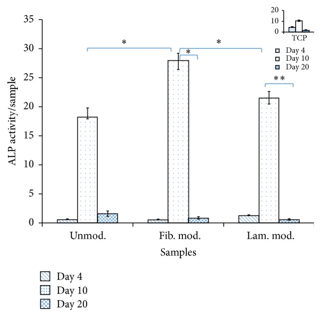
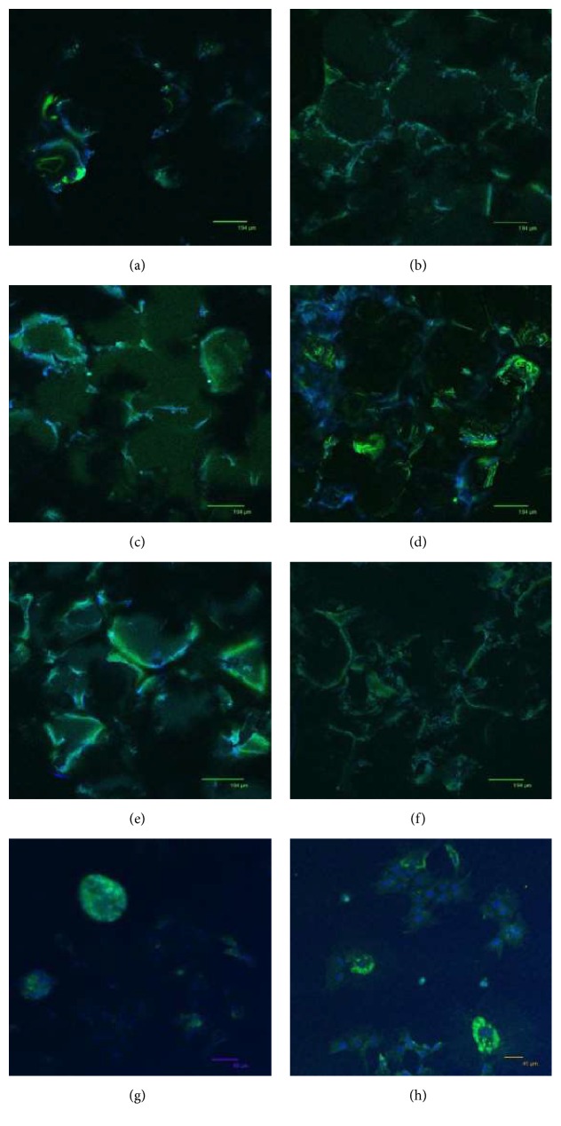
Designing and providing a scaffold are very important for the cells in tissue engineering. Polybutylene succinate (PBS) has high potential as a scaffold for bone regeneration due to its capacity in cell proliferation and differentiation. Also, stem cells from 3rd molar tooth germs were favoured in this study due to their developmentally and replicatively immature nature. In this study, porcine dental germ stem cells (pDGSCs) seeded PBS scaffolds were used to investigate the effects of surface modification with fibronectin or laminin on these scaffolds to improve cell attachment, proliferation, and osteogenic differentiation for tissue engineering applications. The osteogenic potentials of pDGSCs on these modified and unmodified foams were examined to heal bone defects and the effects of fibronectin or laminin modified PBS scaffolds on pDGSC differentiation into bone were compared for the first time. For this study, MTS assay was used to assess the cytotoxic effects of modified and unmodified surfaces. For the characterization of pDGSCs, flow cytometry analysis was carried out. Besides, alkaline phosphatase (ALP) assay, von Kossa staining, real-time PCR, CM-Dil, and immunostaining were applied to analyze osteogenic potentials of pDGSCs. The results of these studies demonstrated that pDGSCs were differentiated into osteogenic cells on fibronectin modified PBS foams better than those on unmodified and laminin modified PBS foams.


Figures

Figure 1
Figure 1
SEM images of PBS scaffolds: (a) 59x, (b) 200x, (c) 500x, and (d) 1,000x objectives.
Figure 2
Figure 2
Degradation of PBS scaffolds with respect to pH throughout 120 days of incubation.
Figure 3
Figure 3
Degradation of PBS scaffolds with respect to weight during 120 days of incubation.
Figure 4
Figure 4
Flow cytometry histogram of pDGSCs with a label by FACSCalibur: (a) only cells without antibody, (b) CD105, (c) CD90, (d) CD44, (e) CD45, and (f) CD34.
Figure 5
Figure 5
Cell proliferation on surface modified and unmodified foams after 3, 4, and 10 days of incubation. Initial cell seeding was 3 × 10⁴ cells/sample (∗∗ p ≤ 0.05). Indent on the top right corner shows the cell proliferation on tissue culture plate.
Figure 6
Figure 6
Normalized ALP activity of pDGSCs on modified and unmodified scaffolds throughout 20 days of incubation ( p ≤ 0.1 and ∗∗ p ≤ 0.05). Indent on the top right corner shows ALP activity of cells seeded on tissue culture plate.
Figure 7
Figure 7
Mineralization of pDGSCs seeded on (a, b) unmodified and fibronectin and laminin modified PBS scaffolds; (c, d) TCP (10x) at the end of (a, c) 10 and (b, d) 20 days of incubation. Arrows indicate mineralized nodules.
Figure 8
Figure 8
Confocal microscopy images of cells seeded on (a, b) unmodified; (c, d) fibronectin modified; and (e, f) laminin modified PBS scaffolds; (g, h) TCP after (a, c, e, g) 10 days and (b, d, f, h) 20 days of incubation. Red stains show actin filaments in the cytoskeleton of the cells stained with Alexa Fluor® 546 Phalloidin and blue stains show the nucleus of the cells stained with TO-PRO-3® Iodide. Scale bars (a) 194 μm, (b) 70 μm, (c) 133 μm, (d) 106 μm, (e) 170 μm, (f) 97 μm, (g) 69 μm, and (h) 186 μm.
Figure 9
Figure 9
Confocal microscopy images of cells seeded on (a, b) unmodified; (c, d) fibronectin modified; and (e, f) laminin modified PBS scaffolds; (g, h) TCP after (a, c, e, g) 10 days and (b, d, f, h) 20 days of incubation. Green stains show the synthesis of collagen in the cytoskeleton of the cells stained with collagen type I and blue stains show the nucleus of the cells stained with TO-PRO-3 Iodide. Scale bars (a, b, c, d, e, f) 194 μm, (g) 85 μm, and (h) 41 μm.
Figure 10
Figure 10
The images of viable cells stained with CellTracker CM-Dil on (a, b) unmodified; (c, d) fibronectin modified; and (e, f) laminin modified PBS scaffolds; (g, h) TCP after 1 day (a, c, e, g) and 7 days (b, d, f, h) of incubation. Scale bars show 100 µm. Arrows indicate viable cells.
Figure 11
Figure 11
Expression levels of (a) alkaline phosphatase (ALP), (b) Runx2, (c) collagen type I (Col I), (d) osteopontin (OPN), and (e) osteocalcin (OCN) on fibronectin (fib.) or laminin (lam.) modified scaffolds, unmodified (unmod.) scaffolds, and TCP (OC refers to “only cell,” i.e., cells seeded on TCP) throughout 20 days of incubation ( p ≤ 0.1 and ∗∗ p ≤ 0.05).

References

    1. Langer R., Vacanti J. P. Tissue engineering. Science. 1993;260(5110):920–926. doi: 10.1126/science.8493529. - DOI - PubMed
    1. Tabata Y. Biomaterial technology for tissue engineering applications. Journal of the Royal Society Interface. 2009;6(3):S311–S324. doi: 10.1098/rsif.2008.0448.focus. - DOI - PMC - PubMed
    1. Ji J., Wang H., Zhang W. Surface modification of biodegradable poly (butylene succinate) by gas PIII. Proceedings of the 3rd International Nanoelectronics Conference (INEC '10); January 2010; Hong Kong. IEEE; pp. 832–833. - DOI
    1. Klee D., Ademovic Z., Bosserhoff A., Hoecker H., Maziolis G., Erli H.-J. Surface modification of poly(vinylidenefluoride) to improve the osteoblast adhesion. Biomaterials. 2003;24(21):3663–3670. doi: 10.1016/S0142-9612(03)00235-7. - DOI - PubMed
    1. Li X., Yao J., Yang X., Tian W., Liu L. Surface modification with fibronectin or collagen to improve the cell adhesion. Applied Surface Science. 2008;255(2):459–461. doi: 10.1016/j.apsusc.2008.06.105. - DOI

LinkOut - more resources