

Clinical and Laboratory Diagnosis of Buruli Ulcer Disease: A Systematic Review
- PMID: 27413382
- PMCID: PMC4931084
- DOI: 10.1155/2016/5310718
Clinical and Laboratory Diagnosis of Buruli Ulcer Disease: A Systematic Review
Abstract
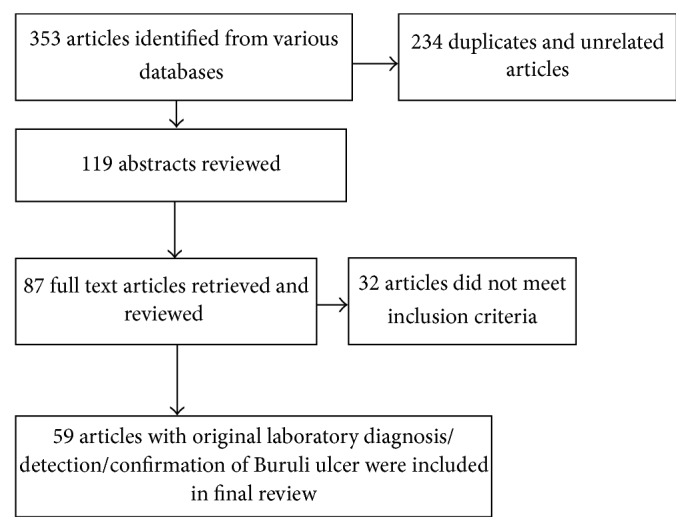
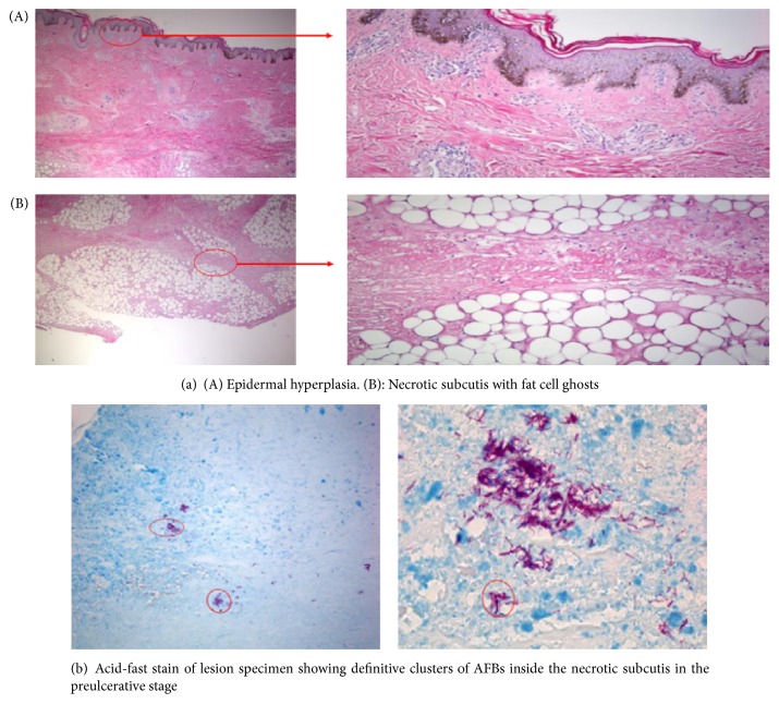
Background. Buruli ulcer (BU) is a necrotizing cutaneous infection caused by Mycobacterium ulcerans. Early diagnosis is crucial to prevent morbid effects and misuse of drugs. We review developments in laboratory diagnosis of BU, discuss limitations of available diagnostic methods, and give a perspective on the potential of using aptamers as point-of-care. Methods. Information for this review was searched through PubMed, web of knowledge, and identified data up to December 2015. References from relevant articles and reports from WHO Annual Meeting of the Global Buruli Ulcer initiative were also used. Finally, 59 articles were used. Results. The main laboratory methods for BU diagnosis are microscopy, culture, PCR, and histopathology. Microscopy and PCR are used routinely for diagnosis. PCR targeting IS2404 is the gold standard for laboratory confirmation. Culture remains the only method that detects viable bacilli, used for diagnosing relapse and accrued isolates for epidemiological investigation as well as monitoring drug resistance. Laboratory confirmation is done at centers distant from endemic communities reducing confirmation to a quality assurance. Conclusions. Current efforts aimed at developing point-of-care diagnostics are saddled with major drawbacks; we, however, postulate that selection of aptamers against MU target can be used as point of care.
Figures
Similar articles
-
An Overview of 10 Years of Activity of a Molecular Laboratory for Buruli Ulcer Diagnosis at a Field Hospital in Benin.J Clin Microbiol. 2023 Jun 20;61(6):e0027423. doi: 10.1128/jcm.00274-23. Epub 2023 May 22. J Clin Microbiol. 2023. PMID: 37212702 Free PMC article.
-
Evaluation of an electricity-independent method for IS2404 Loop-mediated isothermal amplification (LAMP) diagnosis of Buruli ulcer in resource-limited settings.PLoS Negl Trop Dis. 2024 Aug 14;18(8):e0012338. doi: 10.1371/journal.pntd.0012338. eCollection 2024 Aug. PLoS Negl Trop Dis. 2024. PMID: 39141676 Free PMC article.
-
Laboratory diagnosis of Buruli ulcer disease.Future Microbiol. 2010 Mar;5(3):363-70. doi: 10.2217/fmb.10.3. Future Microbiol. 2010. PMID: 20210548 Review.
-
RNA Aptamer That Specifically Binds to Mycolactone and Serves as a Diagnostic Tool for Diagnosis of Buruli Ulcer.PLoS Negl Trop Dis. 2016 Oct 24;10(10):e0004950. doi: 10.1371/journal.pntd.0004950. eCollection 2016 Oct. PLoS Negl Trop Dis. 2016. PMID: 27776120 Free PMC article.
-
PCR detection of Mycobacterium ulcerans-significance for clinical practice and epidemiology.Expert Rev Mol Diagn. 2018 Dec;18(12):1063-1074. doi: 10.1080/14737159.2018.1543592. Epub 2018 Nov 27. Expert Rev Mol Diagn. 2018. PMID: 30381977 Review.
Cited by
-
Analysis of Mycobacterium ulcerans-specific T-cell cytokines for diagnosis of Buruli ulcer disease and as potential indicator for disease progression.PLoS Negl Trop Dis. 2017 Feb 27;11(2):e0005415. doi: 10.1371/journal.pntd.0005415. eCollection 2017 Feb. PLoS Negl Trop Dis. 2017. PMID: 28241036 Free PMC article.
-
A Systematic Review on Suitability of Molecular Techniques for Diagnosis and Research into Infectious Diseases of Concern in Resource-Limited Settings.Curr Issues Mol Biol. 2022 Sep 21;44(10):4367-4385. doi: 10.3390/cimb44100300. Curr Issues Mol Biol. 2022. PMID: 36286015 Free PMC article. Review.
-
Buruli Ulcer: Review of a Neglected Skin Mycobacterial Disease.J Clin Microbiol. 2018 Mar 26;56(4):e01507-17. doi: 10.1128/JCM.01507-17. Print 2018 Apr. J Clin Microbiol. 2018. PMID: 29343539 Free PMC article. Review.
-
Microbiology of secondary infections in Buruli ulcer lesions; implications for therapeutic interventions.BMC Microbiol. 2021 Jan 5;21(1):4. doi: 10.1186/s12866-020-02070-5. BMC Microbiol. 2021. PMID: 33402095 Free PMC article.
-
Diagnostic Accuracy of Clinical and Microbiological Signs in Patients With Skin Lesions Resembling Buruli Ulcer in an Endemic Region.Clin Infect Dis. 2018 Aug 31;67(6):827-834. doi: 10.1093/cid/ciy197. Clin Infect Dis. 2018. PMID: 29538642 Free PMC article.
References
-
- WHO. Buruli ulcer: progress report, 2004–2008. Weekly Epidemiological Record. 2008;83(17):145–154. - PubMed
-
- Ellen D. E., Stienstra Y., Teelken M. A., Dijkstra P. U., Van Der Graaf W. T. A., Van Der Werf T. S. Assessment of functional limitations caused by Mycobacterium ulcerans infection: towards a Buruli ulcer functional limitation score. Tropical Medicine & International Health. 2003;8(1):90–96. doi: 10.1046/j.1365-3156.2003.00976.x. - DOI - PubMed
Publication types
LinkOut - more resources
Full Text Sources
Other Literature Sources
Research Materials
