. 2016 Jul 12:7:13.
doi: 10.1186/s13227-016-0050-x. eCollection 2016.

Conserved expression of vertebrate microvillar gene homologs in choanocytes of freshwater sponges

Affiliations

Conserved expression of vertebrate microvillar gene homologs in choanocytes of freshwater sponges

Jesús F Peña et al. Evodevo. .

Abstract

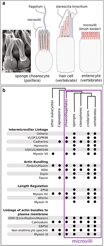
Background: The microvillus is a versatile organelle that serves important functions in disparate animal cell types. However, from a molecular perspective, the microvillus has been well studied in only a few, predominantly vertebrate, contexts. Little is known about how differences in microvillar structure contribute to differences in function, and how these differences evolved. We sequenced the transcriptome of the freshwater sponge, Ephydatia muelleri, and examined the expression of vertebrate microvillar gene homologs in choanocytes-the only microvilli-bearing cell type present in sponges. Sponges offer a distant phylogenetic comparison with vertebrates, and choanocytes are central to discussions about early animal evolution due to their similarity with choanoflagellates, the single-celled sister lineage of modern animals.

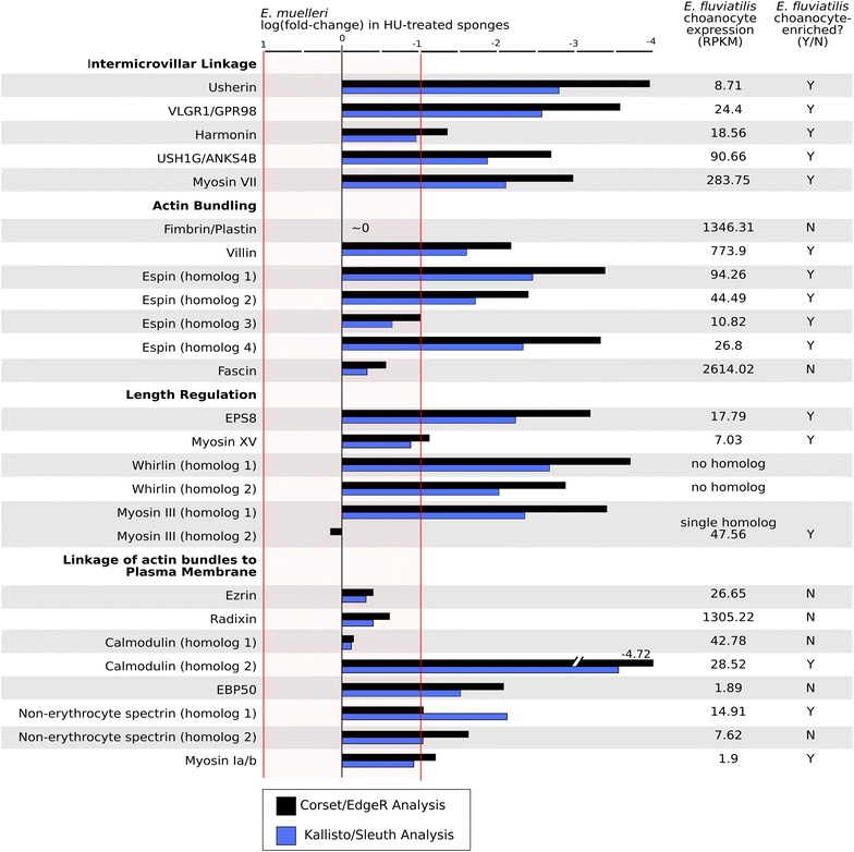
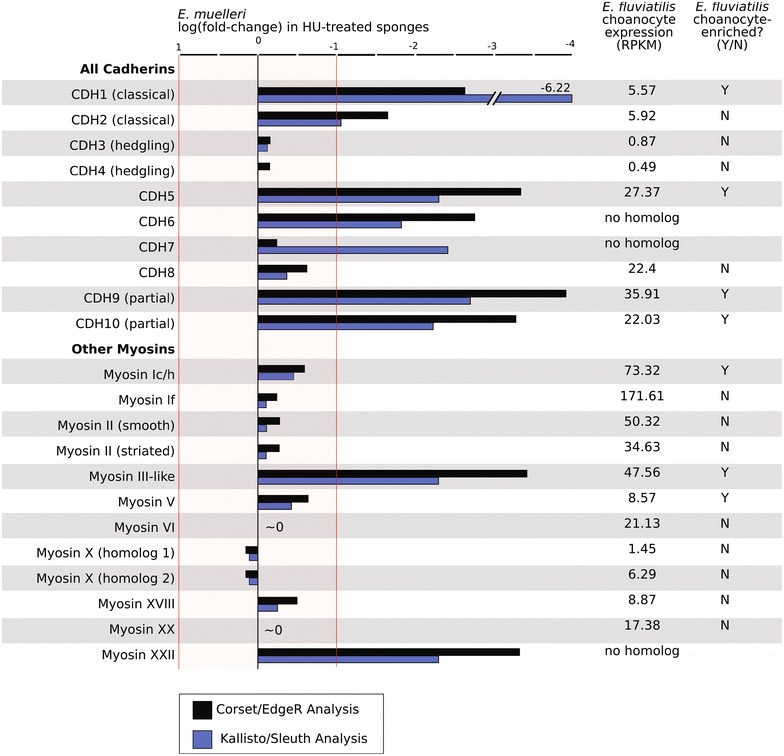
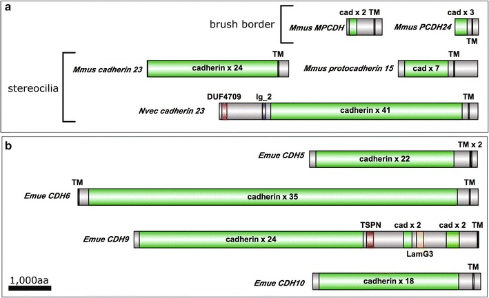
Results: We found that, from a genomic perspective, sponges have conserved homologs of most vertebrate microvillar genes, most of which are expressed in choanocytes, and many of which exhibit choanocyte-specific or choanocyte-enriched expression. Possible exceptions include the cadherins that form intermicrovillar links in the enterocyte brush border and hair cell stereocilia of vertebrates and cnidarians. No obvious orthologs of these proteins were detected in sponges, but at least four candidate cadherins were identified as choanocyte-enriched and might serve this function. In contrast to the evidence for conserved microvillar structure in sponges and vertebrates, we found that choanoflagellates and ctenophores lack homologs of many fundamental microvillar genes, suggesting that microvillar structure may diverge significantly in these lineages, warranting further study.

Conclusions: The available evidence suggests that microvilli evolved early in the prehistory of modern animals and have been repurposed to serve myriad functions in different cellular contexts. Detailed understanding of the sequence by which different microvilli-bearing cell/tissue types diversified will require further study of microvillar composition and development in disparate cell types and lineages. Of particular interest are the microvilli of choanoflagellates, ctenophores, and sponges, which collectively bracket the earliest events in animal evolution.

Keywords: Choanocyte; Ephydatia; Microvilli; Porifera; Sponge; Stereocilia; Transcriptome.


Figures

Fig. 1
Fig. 1
Phylogenetic distribution of microvilli and microvilli-associated proteins. a Sponge choanocytes have an apical ring of actin-cored (red) microvilli that are connected by intermicrovillar links. This structure called a “collar” surrounds a microtubule-cored (blue) flagellum that functions to generate flow through the water-canal system. Microvilli are found in diverse animal cell types. Two of the best-studied examples include mechanosensory hair cells of the vertebrate inner ear, and enterocytes of the vertebrate intestinal epithelium. b Phylogenetic distribution of microvillar proteins that are conserved between sponges and vertebrates
Fig. 2
Fig. 2
Hydroxyurea inhibits choanocyte differentiation. Addition of hydroxyurea (HU) during gemmule hatching, just prior to the differentiation of choanocytes, leads to sponges that lack choanocytes and are enriched for archeocytes, their developmental precursors. a/a’ Low- and high-magnification images a no-treatment control sponge, whereas b/b’ show comparable views of an HU-treated sponge (g gemmule)
Fig. 3
Fig. 3
Choanocyte expression of vertebrate microvillar gene homologs. In E. muelleri, choanocyte expression of candidate microvillar genes was examined through differential expression analysis of HU-treated (i.e., choanocyte-absent) sponges relative to untreated (i.e., choanocyte-present), control sponges. Genes with significantly lower expression in HU-treated sponges (log FC < −1) are interpreted as having elevated expression levels in choanocytes of normal, untreated sponges. In contrast, in E. fluviatilis, choanocyte gene expression levels (RPKM) were determined by direct sequencing of isolated choanocytes and were compared to choanocyte-free cell fractions (summary of expression values is provided in Additional file 2: Supplemental Table 1)
Fig. 4
Fig. 4
Choanocyte expression of cadherins and myosins. Diverse cadherins and myosins regulate the development, structure, and function of microvilli on disparate cell types. We examined choanocyte expression of the full catalog of cadherins and mysoins detected in the E. muelleri transcriptome, and their orthologs identified in the E. fluviatilis transcriptome (summary of expression values is provided in Additional file 2: Supplemental Table 1)
Fig. 5
Fig. 5
Domain architecture of known and candidate intermicrovillar-link cadherins. a Cadherins that link microvilli of the enterocyte brush border, and stereocilia of hair cells of vertebrates, and hair bundles of the cnidarian N. vectensis. b Four cadherins are predicted to have elevated expression levels in choanocytes of E. muelleri. None have obvious orthology with known microvillar-link-forming cadherins, but may function in this capacity (Mmus = Mus musculus, Nvec = Nematostella vectensis, Emue = Ephydatia muelleri, cad = cadherin, TM = transmembrane, DUF = domain of unknown function, TSPN = tetraspanin, LamG3 = Laminin G3)

References

    1. King N. The unicellular ancestry of animal development. Dev Cell. 2004;7(3):313–325. doi: 10.1016/j.devcel.2004.08.010. - DOI - PubMed
    1. Maldonado M. Choanoflagellates, choanocytes, and animal multicellularity. Invertebr Biol. 2004;123(1):1–22. doi: 10.1111/j.1744-7410.2004.tb00138.x. - DOI
    1. Nielsen C. Six major steps in animal evolution: are we derived sponge larvae? Evol Dev. 2008;10(2):241–257. doi: 10.1111/j.1525-142X.2008.00231.x. - DOI - PubMed
    1. Richter DJ, King N. The genomic and cellular foundations of animal origins. Annu Rev Genet. 2013;47:509–537. doi: 10.1146/annurev-genet-111212-133456. - DOI - PubMed
    1. Mah JL, Christensen-Dalsgaard KK, Leys SP. Choanoflagellate and choanocyte collar-flagellar systems and the assumption of homology. Evol Dev. 2014;16(1):25–37. doi: 10.1111/ede.12060. - DOI - PubMed