

Bone Regeneration in Iliac Crestal Defects: An Experimental Study on Sheep
- PMID: 27413746
- PMCID: PMC4931071
- DOI: 10.1155/2016/4086870
Bone Regeneration in Iliac Crestal Defects: An Experimental Study on Sheep
Abstract
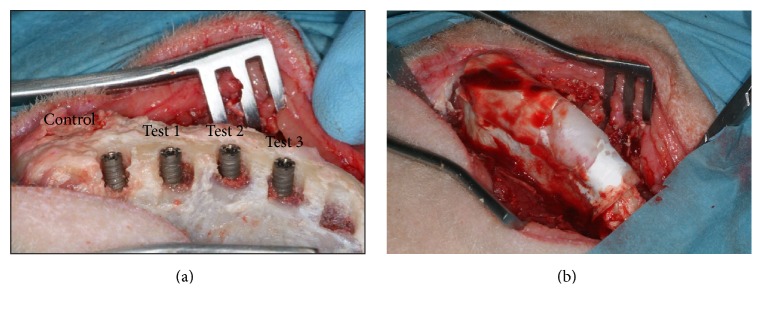
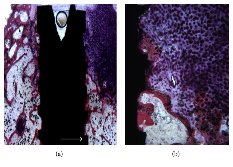
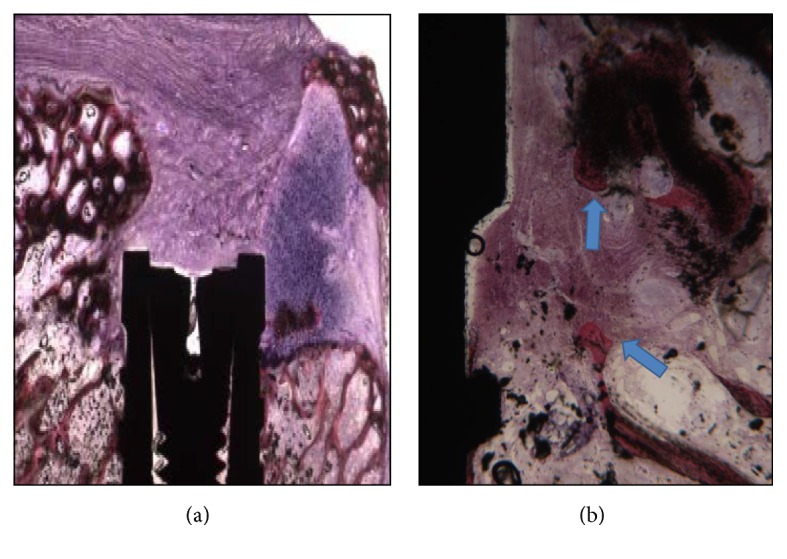
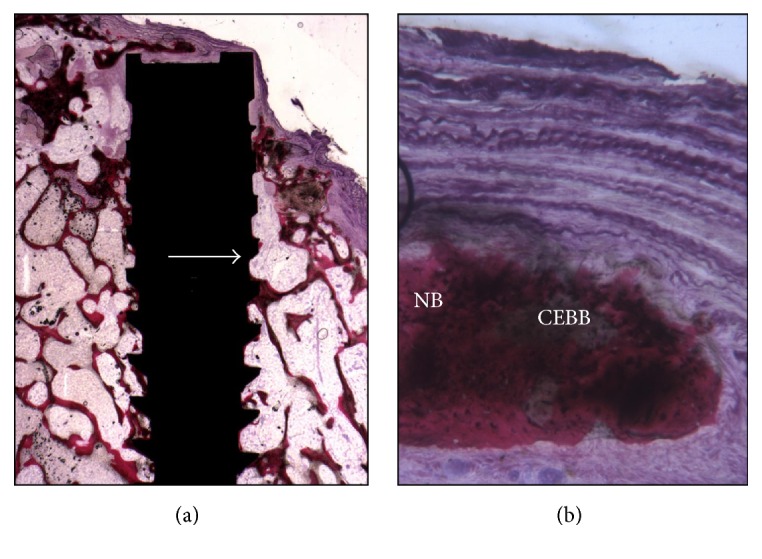
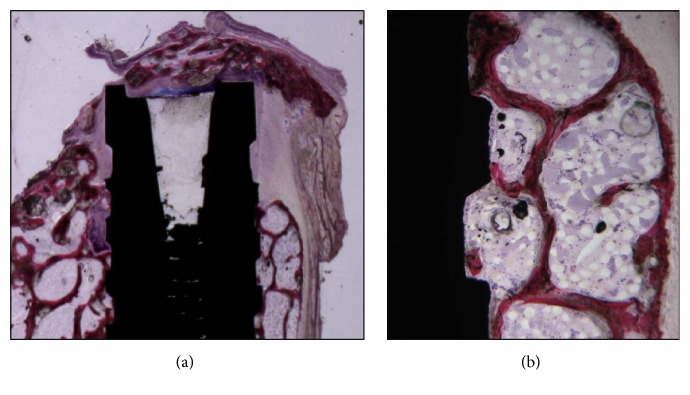
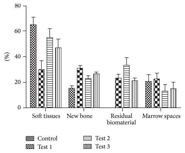
Background. Oral rehabilitation of partially fully edentulous patients with dental implants has become a routine procedure in clinical practice. In a site with a lack of bone GBR is a surgical procedure that provides an augmentation in terms of volume for the insertion of dental implants. Materials and Methods. In the iliac crest of six sheep 4 defects were created where an implant was inserted, three of them with different biomaterials and a control site. All animals were sacrificed after a 4-month healing period. All specimens were processed and analyzed with histomorphometry. Statistical evaluation was done to evaluate percentage of bone defect filled by new bone. Results. All experimental groups showed an increase of the new bone. Higher and highly statistically significant differences were found in the percentages of bone defect filled by new bone in group filled with corticocancellous 250-1000 microns particulate porcine bone mix. Conclusions. This study demonstrates that particulate porcine bone mix and porcine corticocancellous collagenate prehydrated bone mix when used as scaffold are able to induce bone regeneration. Moreover, these data suggest that these biomaterials have higher biocompatibility and are capable of inducing faster and greater bone formation.
Figures
References
-
- Adell R., Eriksson B., Lekholm U., Brånemark P. I., Jemt T. Long-term follow-up study of osseointegrated implants in the treatment of totally edentulous jaws. The International Journal of Oral & Maxillofacial Implants. 1990;5(4):347–359. - PubMed
-
- Albrektsson T., Bergman B., Folmer T. A multicenter study of osseointegrated oral implants. The Journal of Prosthetic Dentistry. 1988;60, article 75 - PubMed
-
- Becker W., Becker B. E., Handlesman M., et al. Bone formation at dehisced dental implant sites treated with implant augmentation material: a pilot study in dogs. The International Journal of Periodontics & Restorative Dentistry. 1990;10(2):92–101. - PubMed
-
- Schenk R. K., Buser D., Hardwick W. R., Dahlin C. Healing pattern of bone regeneration in membrane-protected defects: a histologic study in the canine mandible. The International Journal of Oral & Maxillofacial Implants. 1994;9(1):13–29. - PubMed
Publication types
MeSH terms
Substances
LinkOut - more resources
Full Text Sources
Other Literature Sources
Medical
