. 2016 Jul 14;12(7):e1005746.
doi: 10.1371/journal.ppat.1005746. eCollection 2016 Jul.

Transdifferentiation and Proliferation in Two Distinct Hemocyte Lineages in Drosophila melanogaster Larvae after Wasp Infection

Affiliations

Transdifferentiation and Proliferation in Two Distinct Hemocyte Lineages in Drosophila melanogaster Larvae after Wasp Infection

Ines Anderl et al. PLoS Pathog. .

Abstract

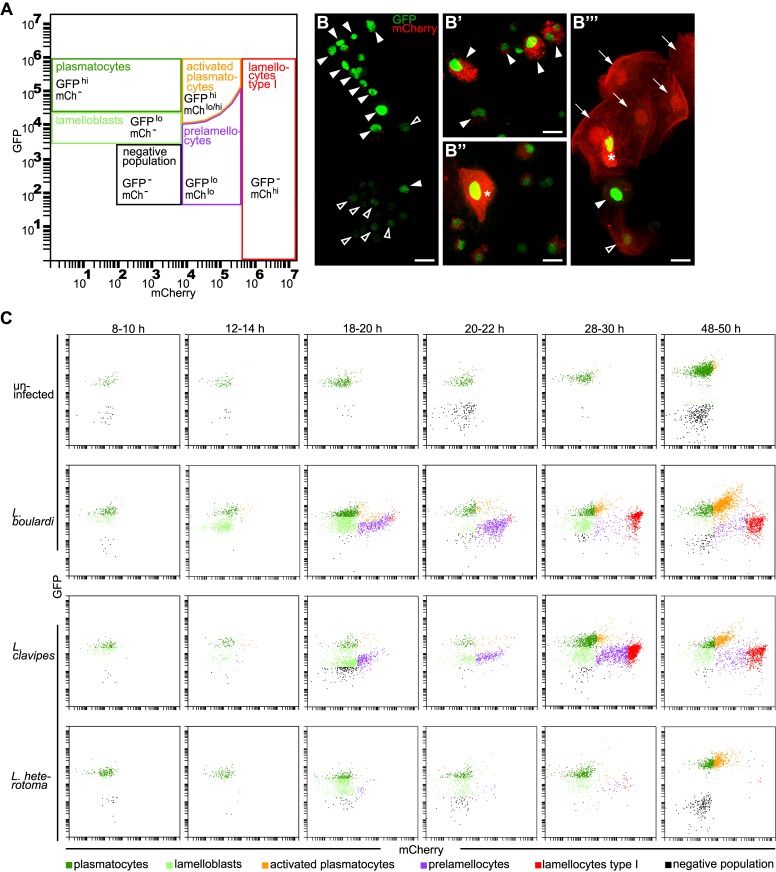
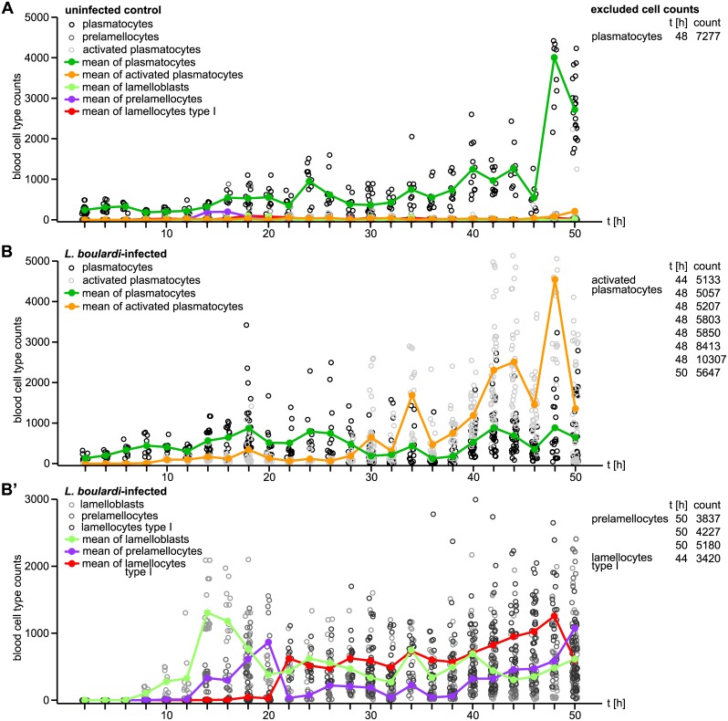
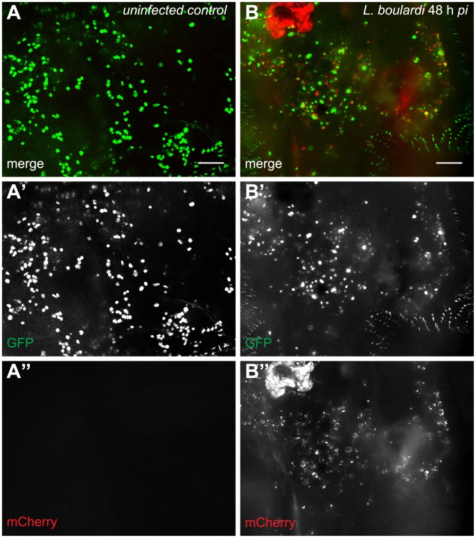
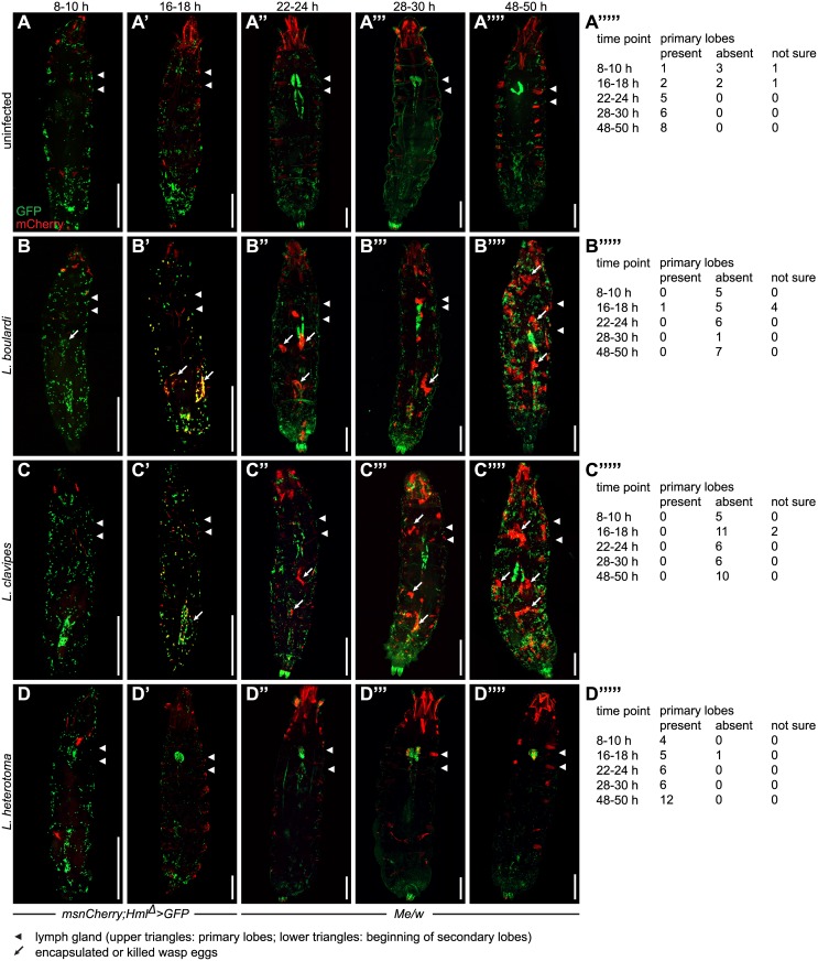
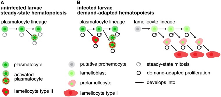
Cellular immune responses require the generation and recruitment of diverse blood cell types that recognize and kill pathogens. In Drosophila melanogaster larvae, immune-inducible lamellocytes participate in recognizing and killing parasitoid wasp eggs. However, the sequence of events required for lamellocyte generation remains controversial. To study the cellular immune system, we developed a flow cytometry approach using in vivo reporters for lamellocytes as well as for plasmatocytes, the main hemocyte type in healthy larvae. We found that two different blood cell lineages, the plasmatocyte and lamellocyte lineages, contribute to the generation of lamellocytes in a demand-adapted hematopoietic process. Plasmatocytes transdifferentiate into lamellocyte-like cells in situ directly on the wasp egg. In parallel, a novel population of infection-induced cells, which we named lamelloblasts, appears in the circulation. Lamelloblasts proliferate vigorously and develop into the major class of circulating lamellocytes. Our data indicate that lamellocyte differentiation upon wasp parasitism is a plastic and dynamic process. Flow cytometry with in vivo hemocyte reporters can be used to study this phenomenon in detail.


Conflict of interest statement

The authors have declared that no competing interests exist.

Figures

Fig 1
Fig 1. Six different hemocyte classes.
(A) Hemocyte gates. Plasmatocytes (GFPhimCh-) and lamelloblasts (GFPlomCh-) expressed only GFP. Activated plasmatocytes (GFPhimChlo) were equal in size to plasmatocytes and had varying amounts of mCherry-positive punctae in their cytoplasm. Similar to activated plasmatocytes, type II lamellocytes expressed both markers (GFPhi mChhi). Type II lamellocytes were larger than plasmatocytes, similar in shape to lamellocytes, and with strong expression of mCherry in their cytoplasm. Prelamellocytes (GFPlomChlo) had low GFP expression while increasing their mCherry expression. Type I lamellocytes (GFP-mChhi) expressed only mCherry. The negative population expressed neither GFP nor mCherry. (B-B”‘) Representative images of hemocyte types. (B) plasmatocytes (filled arrowheads) and lamelloblasts (open arrowheads). (B’) Activated plasmatocytes (filled arrowheads). (B”) Type II lamellocyte (star). (B”‘) Lamellocytes type I (arrows), prelamellocyte (open arrowhead), activated plasmatocyte (filled arrowhead), and lamellocyte type II (star). Scale bars 10 μm. (C) Flow cytometry plots at representative time points after a wasp infection. Me/w larvae were uninfected or infected by L. boulardi, L. clavipes, and L. heterotoma. The time points were chosen to cover major changes in the composition of the hemocyte population in the course of the time line experiment. In uninfected animals mainly plasmatocytes were present in the circulation. The cellular immune response after infection by L. boulardi and L. clavipes proceeded in a stereotypical way. At 10–12 h after infection, two plasmatocyte-like populations, plasmatocytes and lamelloblasts, were present in the circulation. At 18–20 h after infection, lamelloblasts developed into prelamellocytes. Plasmatocytes started to appear already 8–10 h after infection, and were the dominant cell type at 48–50 h. The first type I lamellocytes were seen in the circulation 20–22 h after infection. Large numbers of type I lamellocytes were in the circulation 28–30 h and 48–50 h after infection. A L. heterotoma infection induced a similar immune response until 18–20 h after infection. Then the numbers of prelamellocytes and lamellocytes type I were reduced in comparison to L. boulardi and L. clavipes-infected larvae. 48–50 h after a L. heterotoma infection, plasmatocytes and activated plasmatocytes were the dominant hemocyte types present. They were accompanied by only very few type I lamellocytes.
Fig 2
Fig 2. Timeline of circulating hemocyte counts of age-matched control and wasp-infected larvae.
(A) Total counts of each hemocyte type of uninfected Me/w heterozygous larvae collected every second hour until 50 h after infection. (B) Total counts of plasmatocytes and activated plasmatocytes, as well as (B’) lamelloblasts, prelamellocytes, and type I lamellocytes collected every second hour until 50 h after infection by L. boulardi. The hemocyte population of uninfected control animals consisted mainly of plasmatocytes, whereas the hemocyte populations of infected animals followed a pattern of a demand-adapted hematopoiesis. In order to facilitate plotting, we excluded hemocyte counts of few individual larvae that clearly deviated from the rest. Cell types, time points, and cell counts are presented in tables next to each plot.
Fig 3
Fig 3. Activated plasmatocytes.
Sessile hemocytes of (A-A”) uninfected Me/w control larvae and (B-B”) L. boulardi-infected Me/w larvae 48 h after infection. The images show the terminal segments of two Drosophila larvae. In uninfected controls, plasmatocytes were the predominant sessile cell type, whereas in infected animals activated plasmatocytes were prevalent. The merge and the independent channels are shown separately. Scale bars 50 μm.
Fig 4
Fig 4. Chronological changes in the pattern of sessile cells in Drosophila larvae after a wasp infection.
Sessile hemocyte populations including the lymph glands were imaged in msnCherry;Hml Δ >GFP and Me/w heterozygous control larvae (A-A”“), or at the indicated time points after infection by L. boulardi (B-B”“), L. clavipes (C-C”“), or L. heterotoma (D-D”“). The occurrence of primary lymph gland lobes at indicated time points in uninfected control larvae (A”“‘), L. boulardi-infected (B”“‘), L. clavipes-infected (C”“‘), and L. heterotoma-infected larvae (D”“‘). msnCherry;Hml Δ >GFP larvae were imaged 8–10 h and 16–18 h and Me/w larvae at the remaining time points. While GFP expression in uninfected and infected larvae was restricted to hemocytes, mCherry was also expressed in the pharyngeal muscles, the alary muscles, and in adjacent fibers of the lateral longitudinal muscles of the body wall. This ectopic mCherry expression increased with age. (A-A”“‘). After infection by L. boulardi and L. clavipes, mCherry expression in the lateral longitudinal muscles decreased, but became visible in pericardial cells. No change in ectopic mCherry expression was observed after L. heterotoma infection (B-D”“). In uninfected larvae, sessile plasmatocytes were clearly visible by their GFP expression. The primary and secondary lymph gland lobes were always visible at the later time points, albeit sometimes obscured due to the imaging method (A-A”“). 8–10 h after an infection by L. boulardi and L. clavipes, no obvious change in the pattern of sessile cells had occurred (B, C). The primary lymph gland lobes were never visible (B”“‘, C”“‘). At 16–18 h, fluorescent blood cells outlined the shape of the wasp eggs and started to express mCherry locally on or in the vicinity of the wasp eggs. No systematic loss of sessile blood cells was observed (B’, C’). The primary lymph gland lobes were predominantly absent (B”“‘, C”“‘). At 22–24 h after an infection by L. boulardi and L. clavipes, the mCherry expression became more visible and localized to the wasp eggs, but mCherry-positive cells formed nodules that were not always connected to wasp eggs (B”, C”). Primary lymph gland lobes were always absent while the secondary lobes were visible (B”, C”). At the two final time points after a L. boulardi and L. clavipes infection, total cell numbers had increased. Encapsulated wasp eggs or larvae were clearly visible. In addition, nodules had formed in areas where no wasp eggs or larvae were present. Secondary lymph gland lobes were hypertrophied (B”‘-B”“, C”‘-C”“), whereas primary lymph gland lobes were always absent (B”“‘, C”“‘). A L. heterotoma infection led to a decrease in sessile blood cell numbers. No mCherry-positive nodules were formed during the course of the infection. The primary lymph gland lobes were present and contained mCherry-positive cells starting from 22–24 h. Secondary lymph gland lobes were not visible at the 28–30 h nor at the 48–50 h time points (D-D”“‘). Scale bars 500 μm.
Fig 5
Fig 5. Chronological events during the cellular immune response on the surface of wasp eggs.
Me/w heterozygous Drosophila larvae were infected by (A-D’) L. boulardi, (E-G’) L. clavipes or (H-J) L. heterotoma, and wasp eggs and wasp larvae were dissected and imaged at the indicated time points. L. boulardi eggs were always attached to the gut. Already 8–10 h after infection, plasmatocytes were visible on the wasp eggs and some started to express mCherry, transforming into activated plasmatocytes. The number of plasmatocytes and activated plasmatocytes on the wasp egg increased during the course of infection. In most cases, L. boulardi wasp larvae hatched around 32 h after infection, and some of them were later killed by the cellular immune system. No hemocytes attached to L. clavipes eggs early after infection. Eggs were encapsulated and melanized 28–30 h after infection, and wasp larvae rarely hatched from these eggs. No hemocytes ever attach to the freely floating eggs of L. heterotoma, and the wasp larvae of this species hatched around 38 h after infection.
Fig 6
Fig 6. Proliferation is necessary for the demand-adapted hematopoiesis in Drosophila larvae after a wasp infection.
(A) The experimental design of EdU feeding and subsequent analysis of hemocytes by flow cytometry of age-matched uninfected and infected larvae. Larvae were infected by L. boulardi for two hours, moved to EdU-containing food at the indicated time points and durations, and finally the flow cytometry analysis was carried out accordingly. (B-B”‘) Representative scatter plots of hemocytes of control and wasp-infected larvae after EdU-feeding. EdU was visualized by Alexa647 labeling. (B) EdU feeding 4–8 h and analysis 8 h after infection. In wild-type control larvae approximately half of the hemocytes incorporated EdU, whereas the EdU incorporation rate slightly increased in infected larvae. (B’) EdU-feeding 2–12 h and analysis 12 h after infection. Almost every lamelloblast, plasmatocyte, and activated plasmatocyte incorporated EdU. (B”) EdU-feeding: 4–8 h and analysis 28 h after infection. Plasmatocytes, activated plasmatocytes, lamelloblasts, prelamellocytes and lamellocytes are shown separately. The amount of EdU incorporating plasmatocyte types after infection approximately equaled that of controls, whereas many of the infection-induced prelamellocytes and lamellocytes incorporated EdU. (B”‘) EdU feeding: 28–32 h and analysis 48 h after infection. Plasmatocytes, activated plasmatocytes, lamelloblasts, prelamellocytes and lamellocytes are shown separately. Slightly more plasmatocytes and lamelloblasts of infected larvae were EdU-positive, while the number of EdU-positive activated plasmatocytes, prelamellocytes and lamellocytes was again high. (C) Percentages of EdU-positive total hemocyte numbers of wasp-infected and control larvae. (D) Quantification of EdU incorporation experiments in (B-B”‘) according to hemocyte types. One to three replicates were conducted for each EdU- feeding scheme. pc—plasmatocytes.
Fig 7
Fig 7. All larval hemocyte types are able to divide, except for lamellocytes.
(A-A”‘) Hemocytes were able to divide on wasp eggs. Arrowheads point to an example of a dividing activated plasmatocyte and a dividing plasmatocyte. Scale bars 50 μm. (B-B’) Sessile blood cells divided in control and L. boulardi-infected larvae 48 h after infection. eaterDsRed;Hml Δ > was crossed with w;He>S/G2/M-Green to obtain eaterDsRed;Hml Δ >;He>S/G2/M-Green. Observe that due to the genotype only plasmatocytes are visible after infection! The number of sessile cells increased after a wasp infection. In the wasp-infected larva, the primary lymph gland lobes were no longer present, whereas they were still visible in the control larva. Arrowheads point to the locations of the primary lymph gland lobes. Scale bars 100 μm. (C) Quantification of dividing cell types after EdU feeding 4–8 h and analysis 28 h after infection of uninfected and L. boulardi-infected larvae. Hemocytes of offspring of crosses of msnCherry;eaterDsRed,Hml Δ > to w;He>S/G2/M-Green were imaged and analysed for the incorporation of EdU-Alexa647, and/or the expression of S/G2/M-Green. The total numbers of evaluated cells per cell type are indicated above the bar plots of each hemocyte type (pc: plasmatocytes). All cell types incorporated EdU. All cell types but lamellocytes expressed Green. All cell types except for lamellocytes incorporated EdU and simultaneously expressed >S/G2/M-Green. The gating strategy is explained in S9 Fig. (D-E”“‘) Representative images of cells analyzed in (C). Arrowheads point to lamellocytes. None of the lamellocytes expressed Green (E”“), but one lamellocyte that had incorporated EdU is shown (E”‘). Scale bars 50 μm.
Fig 8
Fig 8. Activation of plasmatocytes on wasp eggs.
(A-C) Still images of hemocytes on the surface of a wasp egg, taken from a time lapse experiment of Me/w larvae infected by L. boulardi. Wasp eggs with attached hemocytes were dissected out 14 h after infection and the attached hemocytes are shown at representative time points after infection: (A) 14 h, (B) 18:35 h and (C) 26:55 h. Plasmatocytes started to express and to continuously increase their expression levels of mCherry, spread on the wasp egg, and thereby transformed into type II lamellocytes. The corresponding time-lapse video can be found at S1 Video. Scale bars 50 μm.
Fig 9
Fig 9. Suppression of edin expression in the fat body inhibits the formation of lamelloblasts.
(A) Knock-down of edin in the fat body reduced total hemocyte numbers 14 h after infection by L. boulardi (ANOVA: replicate*genotype: F = 1.983, p = 0.121, df = 3; replicate: F = 0.746, p = 0.39, df = 1; genotype: F = 10.923, p < 0.001, df = 3; for pairwise comparisons we used Tukey`s HSD). Three independent experiments are shown for the genotypes Me, Me;Fb>edin, and Me;Fb>edin KK, and two for the genotype Me;edin. Hemocyte numbers of each experiment are represented by dots in different shades and the mean hemocyte numbers by red bars. At least ten larvae were bled per genotype and experiment. (B) Knock-down of edin in the fat body reduced the number of lamelloblasts (Kruskal-Wallis rank sum test: genotype: H = 44.913, p < 0.001, df = 3; for pairwise comparisons we used the independent two-group Mann-Whitney U Test) 14 h after infection by L. boulardi. Identical experiments as in (A), but the cell numbers for each genotype are plotted separately. The mean cell numbers for each cell type are plotted as colored bars. ns—not significant.
Fig 10
Fig 10. Antibody staining of hemocytes fixed on glass slides and on L. boulardi eggs.
(A) Staining of Me-expressing hemocytes of age-matched control and L. boulardi-infected larvae with NimC1/P1, Atilla/L1, L2, Myospheroid/L4, and L6 antibodies at 8, 18, 20, 28, and 48h after infection. Individual hemocyte types are shown in separate graphs. Hemocytes of uninfected control larvae were primarily plasmatocytes and stained mainly with NimC1/P1 antibody at all time points. All blood cells of infected larvae stained with NimC1/P1 as long as they expressed eaterGFP (> 75% in all cell types). The percentage of expression of NimC1 was lowest in lamelloblasts from 20 h after infection onwards. Lamellocytes never expressed NimC1 and expressed all of the lamellocyte antigens. After infection, all blood cell types stained to some extent with lamellocyte markers. The L4 antigen was the first and the L6 antigen the last to be expressed by circulating hemocytes. Some cell types were rare at certain time points. A missing data point indicates that fewer than ten cells were counted, and these data were excluded from the analysis. (B-D”‘) Hemocytes of Me/w on L. boulardi eggs stained with (B-B”‘) NimC1/P1, (C-C”‘) L4, and (D-D”‘) Atilla/L1 14 h after infection. Alexa405 was used as the secondary antibody label. All channels are shown separately and as a merge. Scale bars 50 μm. The NimC1/P1 antibody did not recognize lamellocytes on the wasp egg, but did recognize plasmatocytes and activated plasmatocytes. Myospheroid/L4 and Attila/L1 stained most plasmatocytes and activated plasmatocytes on the wasp egg.
Fig 11
Fig 11. Model.
(A) Steady-state hematopoiesis: Hemocytes of the plasmatocyte lineage prevail. Plasmatocytes self-renew, proliferate, and give rise to a small percentage of activated plasmatocytes. (B) Demand-adapted hematopoiesis. After wasp infection, hemocytes of the plasmatocyte lineage increase their proliferation rate. Plasmatocytes give rise to activated plasmatocytes with large and abundant mCherry-positive foci. And plasmatocytes transdifferentiate into type II lamellocytes on the wasp egg. A wasp infection induces the lamellocytes lineage. Putative prohemocytes that reside within the sessile hemocyte bands, and which are double-negative for the dual reporter constructs, give rise to circulating vigorously dividing lamelloblasts that transiently express GFP. These cells develop into type I lamellocytes via an intermediate prelamellocyte. All cell types are able to divide with the exception of type I lamellocytes.

Similar articles

Cited by

References

    1. Evans CJ, Hartenstein V, Banerjee U. Thicker than blood: conserved mechanisms in Drosophila and vertebrate hematopoiesis. Dev Cell. 2003; 5: 673–690. - PubMed
    1. Williams MJ. Drosophila hemopoiesis and cellular immunity. J Immunol. 2007; 178: 4711–4716. - PubMed
    1. Krzemien J, Crozatier M, Vincent A. Ontogeny of the Drosophila larval hematopoietic organ, hemocyte homeostasis and the dedicated cellular immune response to parasitism. Int J Dev Biol. 2010; 54: 1117–1125. 10.1387/ijdb.093053jk - DOI - PubMed
    1. Honti V, Csordás G, Kurucz E, Márkus R, Andó I. The cell-mediated immunity of Drosophila melanogaster: hemocyte lineages, immune compartments, microanatomy and regulation. Dev Comp Immunol. 2014; 42: 47–56. 10.1016/j.dci.2013.06.005 - DOI - PubMed
    1. Dudzic JP, Kondo S, Ueda R, Bergman CM, Lemaitre B. Drosophila innate immunity: regional and functional specialization of prophenoloxidases. BMC Biol. 2015; 13: 81 10.1186/s12915-015-0193-6 - DOI - PMC - PubMed

Publication types

LinkOut - more resources