. 2016 Jul 14;11(7):e0158969.
doi: 10.1371/journal.pone.0158969. eCollection 2016.

Local Epidermal Growth Factor Receptor Signaling Mediates the Systemic Pathogenic Effects of Staphylococcus aureus Toxic Shock Syndrome

Affiliations

Local Epidermal Growth Factor Receptor Signaling Mediates the Systemic Pathogenic Effects of Staphylococcus aureus Toxic Shock Syndrome

Laura M Breshears et al. PLoS One. .

Abstract

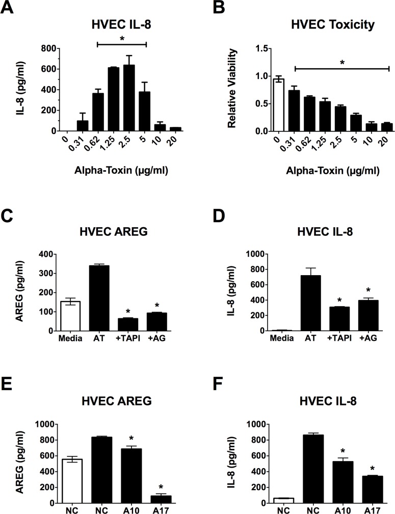
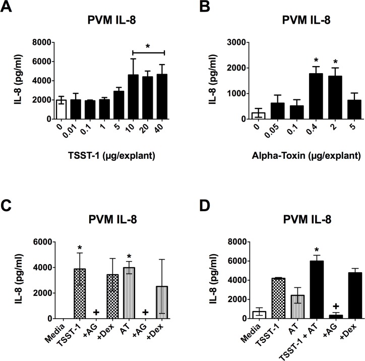
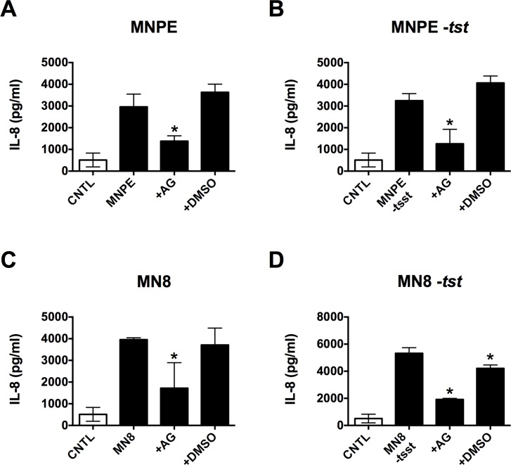
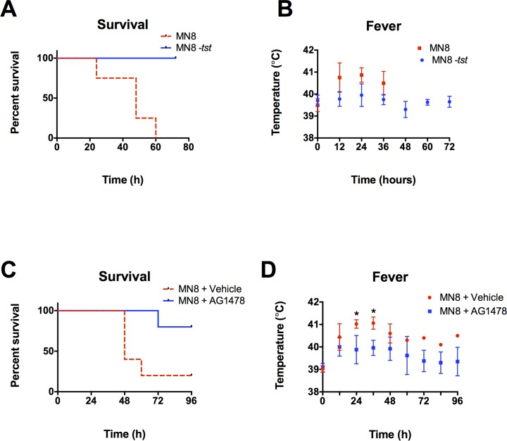
Secreted factors of Staphylococcus aureus can activate host signaling from the epidermal growth factor receptor (EGFR). The superantigen toxic shock syndrome toxin-1 (TSST-1) contributes to mucosal cytokine production through a disintegrin and metalloproteinase (ADAM)-mediated shedding of EGFR ligands and subsequent EGFR activation. The secreted hemolysin, α-toxin, can also induce EGFR signaling and directly interacts with ADAM10, a sheddase of EGFR ligands. The current work explores the role of EGFR signaling in menstrual toxic shock syndrome (mTSS), a disease mediated by TSST-1. The data presented show that TSST-1 and α-toxin induce ADAM- and EGFR-dependent cytokine production from human vaginal epithelial cells. TSST-1 and α-toxin also induce cytokine production from an ex vivo porcine vaginal mucosa (PVM) model. EGFR signaling is responsible for the majority of IL-8 production from PVM in response to secreted toxins and live S. aureus. Finally, data are presented demonstrating that inhibition of EGFR signaling with the EGFR-specific tyrosine kinase inhibitor AG1478 significantly increases survival in a rabbit model of mTSS. These data indicate that EGFR signaling is critical for progression of an S. aureus exotoxin-mediated disease and may represent an attractive host target for therapeutics.


Conflict of interest statement

Competing Interests: The authors have declared that no competing interests exist.

Figures

Fig 1
Fig 1. α-toxin induces AREG shedding and IL-8 production from HVECs.
HVECs were exposed to α-toxin for 6 h and then processed for toxicity via MTT assay or IL-8 secretion and AREG shedding via ELISA. Where inhibitors were used, they were applied to HVECs 30 min prior to addition of α-toxin. (A) IL-8 secretion from and (B) viability of HVECs exposed to various doses of α-toxin. For both curves, the asterisks indicate doses showing significant differences from 0 (p < 0.0001). (C) AREG shedding and (D) IL-8 secretion in response to α-toxin is dampened in the presence of TAPI-1 and AG1478. White bars indicate media alone, black bars represent α-toxin treatment at 1 μg/ml. Asterisks indicate significant differences from α-toxin (AT) alone (p < 0.0001). (E) AREG shedding and (F) IL-8 secretion in response to α-toxin are dampened in ADAM10 (A10) and ADAM17 (A17) KD cell lines. White bars indicate media alone on negative control (NC) siRNA cells, black bars represent α-toxin treatment at 1 μg/ml. Asterisks indicate significant differences from α-toxin treated NC cells (p < 0.0001).
Fig 2
Fig 2. IL-8 production from PVM in response to TSST-1 and α-toxin is dependent on EGFR signaling.
PVM explants were exposed to TSST-1 and/or α-toxin for 6 h and then processed for IL-8 production via ELISA. Where inhibitors were used, they were applied to explants 30 minutes prior to addition of toxin(s). IL-8 is produced in response to both (A) TSST-1 and (B) α-toxin in a dose-dependent manner. For both curves, the asterisks indicate doses showing significant differences from 0 (p < 0.0004). (C) IL-8 production in response to high doses of both TSST-1 (20 μg/explant) and α-toxin (AT) (2 μg/explant) is completely abrogated in the presence of AG1478 (AG– 40 μg/explant), but the dextrin vehicle (Dex– 10 μl of 15%) alone has no affect. Checkered bars represent TSST-1 treatment and striped bars represent AT treatment. Asterisks indicate significant differences from media alone, while crosses indicate significant differences from toxin alone (p < 0.0003). (D) Low doses of TSST-1 (5 μg/explant) and AT (25 ng/explant) have an additive effect on IL-8 production that is reduced to basal levels in the presence of AG1478 (4 μg/explant) with no dextrin vehicle effect (10 μl of 15%). White bars indicate media alone, checkered bars represent TSST-1 treatment, striped bars represent AT treatment, black bars represent TSST-1 + AT treatment. Asterisk indicates significant difference from media, TSST-1 and AT alone (p < 0.03), while cross indicates significant difference from TSST-1 + AT (p < 0.0009).
Fig 3
Fig 3. EGFR signaling mediates the PVM IL-8 response to S. aureus.
PVM explants were inoculated with ~ 107 CFU/explant and incubated for 6 h ± 4 μg/explant AG1478 (AG) or 4 μl/explant 10% DMSO vehicle prior to processing for IL-8 production (via ELISA) and CFU determination. (A-E) White bar, uninfected control (CNTL), black bars indicate presence of bacteria. Incubation of PVM with AG prior to infection with (A) MNPE, (B) MNPE -tstH, (C) MN8, or (D) MN8 -tstH significantly reduced IL-8 production (asterisks, p < 0.0002). DMSO alone had no effect.
Fig 4
Fig 4. EGFR signaling is required for mTSS progression in vivo.
(A, B) WT MN8 or MN8 -tstH were administered at 5 x 10⁸ twice daily for 3 days or until death, N = 1, n = 4. (A) Survival is significantly increased (p < 0.0069) and (B) fever is generally reduced in animals challenged with MN8 -tstH versus WT MN8. (C, D) In separate experiments, rabbits were intravaginally challenged with ~ 1010 MN8 + 8 mg/ml AG1478 or 30% beta cyclodextrin vehicle twice daily for 4 days or until death, N = 1, n = 5. (C) Survival is significantly increased in animals treated with AG1478 (p < 0.03). (D) Fever is generally decreased in animals treated with AG1478 reaching significance at 24 and 36 h, just prior to the death of the majority of infected, untreated animals (p < 0.005).

References

    1. Huovila A-PJ, Turner AJ, Pelto-Huikko M, Kärkkäinen I, Ortiz RM. Shedding light on ADAM metalloproteinases. Trends Biochem Sci. 2005;30:413–422. - PubMed
    1. Oda K, Matsuoka Y, Funahashi A, Kitano H. A comprehensive pathway map of epidermal growth factor receptor signaling. Mol Syst Biol. 2005;1:E1–E17. - PMC - PubMed
    1. Roupé KM, Nybo M, Sjöbring U, Alberius P, Schmidtchen A, Sørensen OE. Injury is a major inducer of epidermal innate immune responses during wound healing. J Invest Dermatol. 2010;130:1167–1177. 10.1038/jid.2009.284 - DOI - PubMed
    1. Tran QT, Kennedy LH, Leon Carrion S, Bodreddigari S, Goodwin SB, Sutter CH, et al. EGFR regulation of epidermal barrier function. Physiol Genomics. 2012;44:455–469. 10.1152/physiolgenomics.00176.2011 - DOI - PMC - PubMed
    1. Pastore S, Mascia F, Mariani V, Girolomoni G. The epidermal growth factor receptor system in skin repair and inflammation. J Invest Dermatol. 2008;128:1365–1374. - PubMed