

Extracellular matrix composition defines an ultra-high-risk group of neuroblastoma within the high-risk patient cohort
- PMID: 27415013
- PMCID: PMC4985353
- DOI: 10.1038/bjc.2016.210
Extracellular matrix composition defines an ultra-high-risk group of neuroblastoma within the high-risk patient cohort
Abstract
Background: Although survival for neuroblastoma patients has dramatically improved in recent years, a substantial number of children in the high-risk subgroup still die.
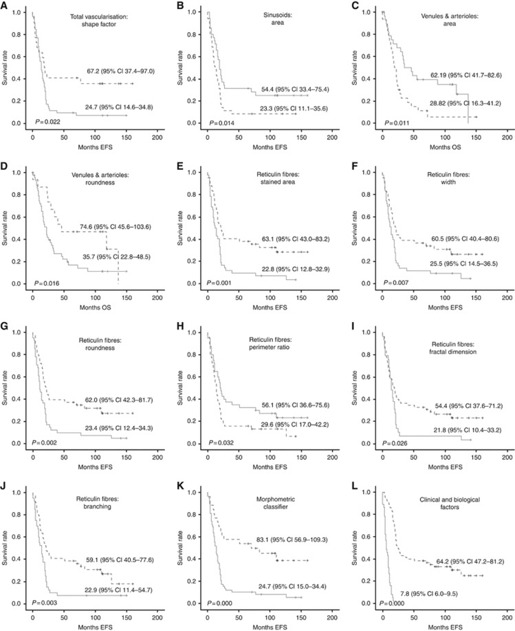
Methods: We aimed to define a subgroup of ultra-high-risk patients from within the high-risk cohort. We used advanced morphometric approaches to quantify and characterise blood vessels, reticulin fibre networks, collagen type I bundles, elastic fibres and glycosaminoglycans in 102 high-risk neuroblastomas specimens. The Kaplan-Meier method was used to correlate the analysed elements with survival.
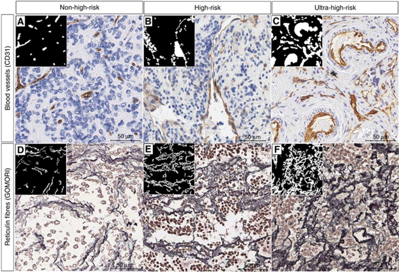
Results: The organisation of blood vessels and reticulin fibres in neuroblastic tumours defined an ultra-high-risk patient subgroup with 5-year survival rate <15%. Specifically, tumours with irregularly shaped blood vessels, large sinusoid-like vessels, smaller and tortuous venules and arterioles and with large areas of reticulin fibres forming large, crosslinking, branching and haphazardly arranged networks were linked to the ultra-high-risk phenotype.
Conclusions: We demonstrate that quantification of tumour stroma components by morphometric techniques has the potential to improve risk stratification of neuroblastoma patients.
Figures
Similar articles
-
A stiff extracellular matrix is associated with malignancy in peripheral neuroblastic tumors.Pediatr Blood Cancer. 2017 Sep;64(9). doi: 10.1002/pbc.26449. Epub 2017 Jan 25. Pediatr Blood Cancer. 2017. PMID: 28121069
-
1p36 deletion results in a decrease in glycosaminoglycans which is associated with aggressiveness in neuroblastic tumors.Histol Histopathol. 2018 May;33(5):487-495. doi: 10.14670/HH-11-947. Epub 2017 Nov 23. Histol Histopathol. 2018. PMID: 29168879
-
Differential distribution of the fibres of the collagenous and elastic systems and of glycosaminoglycans in the rat pubic joint.J Submicrosc Cytol Pathol. 2001 Oct;33(4):463-72. J Submicrosc Cytol Pathol. 2001. PMID: 11989780
-
Progress towards personalized therapeutics: biologic- and risk-directed therapy for neuroblastoma.Expert Rev Neurother. 2011 Oct;11(10):1411-23. doi: 10.1586/ern.11.103. Expert Rev Neurother. 2011. PMID: 21955198 Review.
-
Structural biology of the fibres of the collagenous and elastic systems.Cell Biol Int. 1996 Jan;20(1):15-27. doi: 10.1006/cbir.1996.0004. Cell Biol Int. 1996. PMID: 8936403 Review.
Cited by
-
Clinically relevant treatment of PDX models reveals patterns of neuroblastoma chemoresistance.Sci Adv. 2022 Oct 28;8(43):eabq4617. doi: 10.1126/sciadv.abq4617. Epub 2022 Oct 28. Sci Adv. 2022. PMID: 36306349 Free PMC article.
-
Targeting the Tumor Microenvironment in Neuroblastoma: Recent Advances and Future Directions.Cancers (Basel). 2020 Jul 25;12(8):2057. doi: 10.3390/cancers12082057. Cancers (Basel). 2020. PMID: 32722460 Free PMC article. Review.
-
Improved risk stratification by PET-based intratumor heterogeneity in children with high-risk neuroblastoma.Front Oncol. 2022 Oct 24;12:896593. doi: 10.3389/fonc.2022.896593. eCollection 2022. Front Oncol. 2022. PMID: 36353561 Free PMC article.
-
A three-dimensional bioprinted model to evaluate the effect of stiffness on neuroblastoma cell cluster dynamics and behavior.Sci Rep. 2020 Apr 14;10(1):6370. doi: 10.1038/s41598-020-62986-w. Sci Rep. 2020. PMID: 32286364 Free PMC article.
-
Emerging Neuroblastoma 3D In Vitro Models for Pre-Clinical Assessments.Front Immunol. 2020 Nov 26;11:584214. doi: 10.3389/fimmu.2020.584214. eCollection 2020. Front Immunol. 2020. PMID: 33324402 Free PMC article. Review.
References
-
- Afratis N, Gialeli C, Nikitovic D, Tsegenidis T, Karousou E, Theocharis AD, Pavao MS, Tzanakakis GN, Karamanos NK (2012) Glycosaminoglycans: key players in cancer cell biology and treatment. FEBS J 279: 1177–1197. - PubMed
-
- Ambros IM, Benard J, Boavida M, Bown N, Caron H, Combaret V, Couturier J, Darnfors C, Delattre O, Freeman-Edward J, Gambini C, Gross N, Hattinger CM, Luegmayr A, Lunec J, Martinsson T, Mazzocco K, Navarro S, Noguera R, O'Neill S, Potschger U, Rumpler S, Speleman F, Tonini GP, Valent A, Van Roy N, Amann G, De Bernardi B, Kogner P, Ladenstein R, Michon J, Pearson AD, Ambros PF (2003) Quality assessment of genetic markers used for therapy stratification. J Clin Oncol 21: 2077–2084. - PubMed
-
- Ambros PF, Ambros IM, Brodeur GM, Haber M, Khan J, Nakagawara A, Schleiermacher G, Speleman F, Spitz R, London WB, Cohn SL, Pearson AD, Maris JM (2009) International consensus for neuroblastoma molecular diagnostics: report from the International Neuroblastoma Risk Group (INRG) Biology Committee. Br J Cancer 100: 1471–1482. - PMC - PubMed
-
- Ariffin AB, Forde PF, Jahangeer S, Soden DM, Hinchion J (2014) Releasing pressure in tumors: what do we know so far and where do we go from here? A review. Cancer Res 74: 2655–2662. - PubMed
-
- Barry-Hamilton V, Spangler R, Marshall D, McCauley S, Rodriguez HM, Oyasu M, Mikels A, Vaysberg M, Ghermazien H, Wai C, Garcia CA, Velayo AC, Jorgensen B, Biermann D, Tsai D, Green J, Zaffryar-Eilot S, Holzer A, Ogg S, Thai D, Neufeld G, Van Vlasselaer P, Smith V (2010) Allosteric inhibition of lysyl oxidase-like-2 impedes the development of a pathologic microenvironment. Nat Med 16: 1009–1017. - PubMed
Publication types
MeSH terms
Substances
LinkOut - more resources
Full Text Sources
Other Literature Sources
Medical
