. 2016 Jun;7(1):10.1128/ecosalplus.ESP-0021-2015.
doi: 10.1128/ecosalplus.ESP-0021-2015.

C4-Dicarboxylate Utilization in Aerobic and Anaerobic Growth

Affiliations

C4-Dicarboxylate Utilization in Aerobic and Anaerobic Growth

Gottfried Unden et al. EcoSal Plus. 2016 Jun.

Abstract

C4-dicarboxylates and the C4-dicarboxylic amino acid l-aspartate support aerobic and anaerobic growth of Escherichia coli and related bacteria. In aerobic growth, succinate, fumarate, D- and L-malate, L-aspartate, and L-tartrate are metabolized by the citric acid cycle and associated reactions. Because of the interruption of the citric acid cycle under anaerobic conditions, anaerobic metabolism of C4-dicarboxylates depends on fumarate reduction to succinate (fumarate respiration). In some related bacteria (e.g., Klebsiella), utilization of C4-dicarboxylates, such as tartrate, is independent of fumarate respiration and uses a Na+-dependent membrane-bound oxaloacetate decarboxylase. Uptake of the C4-dicarboxylates into the bacteria (and anaerobic export of succinate) is achieved under aerobic and anaerobic conditions by different sets of secondary transporters. Expression of the genes for C4-dicarboxylate metabolism is induced in the presence of external C4-dicarboxylates by the membrane-bound DcuS-DcuR two-component system. Noncommon C4-dicarboxylates like l-tartrate or D-malate are perceived by cytoplasmic one-component sensors/transcriptional regulators. This article describes the pathways of aerobic and anaerobic C4-dicarboxylate metabolism and their regulation. The citric acid cycle, fumarate respiration, and fumarate reductase are covered in other articles and discussed here only in the context of C4-dicarboxylate metabolism. Recent aspects of C4-dicarboxylate metabolism like transport, sensing, and regulation will be treated in more detail. This article is an updated version of an article published in 2004 in EcoSal Plus. The update includes new literature, but, in particular, the sections on the metabolism of noncommon C4-dicarboxylates and their regulation, on the DcuS-DcuR regulatory system, and on succinate production by engineered E. coli are largely revised or new.


Figures

Figure 1
Figure 1
Different strategies for aerobic (A) and anaerobic (B) utilization of C4-dicarboxylates by E. coli using the tricarboxylic acid cycle (TCA) (A) and fumarate respiration (B). Anaerobic utilization of L-tartrate and citrate by Salmonella and Klebsiella occurs by a different route that involves conversion of OAA (oxaloacetate) to pyruvate by a Na+-dependent oxaloacetate decarboxylase (without fumarate respiration).
Figure 2
Figure 2
Pathways of aerobic C4-dicarboxylate utilization by E. coli. The scheme gives the sequence of enzyme reactions for the utilization of succinate (Succ) and other C4-dicarboxylates in aerobic growth (1 succinate + 3.5 O2→ 4 CO2 + 3 H2O). For simplicity, not all intermediates for the conversion of citrate to succinate, and of PEP to pyruvate (Pyr), are shown. Membrane-associated or integral enzymes are indicated by their locations. DctA, SatP, and DauA are transporters for the uptake of the common C4-dicarboxylates at pH > 6, pH 6, and pH 5, respectively. DctA catalyzes also the uptake of the noncommon C4-dicarboxylates D-malate and L-tartrate under aerobic conditions. DcuA is produced constitutively and transports C4-dicarboxylates and particularly L-aspartate (L-Asp). The pathway for aerobic oxidation of L-tartrate is not known. AcCoA, acetyl-CoA; CS, citrate synthase; DctA, aerobic C4-dicarboxylate transporter; DmlA, D-malate dehydrogenase; Sdh, succinate dehydrogenase SdhABCD; FumA and FumC (aerobic) fumarase; Mal, malate; Mdh, cytosolic (NADH-dependent) malate dehydrogenase; Mqo, membrane-associated malate-quinone oxidoreductase; MaeB and SfcA, NAD(P)H-dependent malic enzymes; OAA, oxaloacetic acid; Pck, PEP carboxykinase; PDH, pyruvate dehydrogenase; TCA, tricarboxylic acid cycle. For details, see text.
Figure 3
Figure 3
Anaerobic catabolic reactions of common C4-dicarboxylates (fumarate, l-malate, aspartate) and relation to fumarate respiration (A) and use of aspartate for anabolic reactions (B). (A) The scheme shows the pathways for the uptake of the C4-dicarboxylates, conversion to fumarate, and formation of succinate by fumarate reduction. From the fumarate reductase system, only fumarate reductase is given; details of fumarate respiration are shown in Fig. 4. DcuA is shown to function in L-Asp/Succ antiport where L-Asp utilization is linked to fumarate respiration (right part). (B) The scheme shows the role of DcuA in the constitutive uptake of L-Asp (or other C4-dicarboxylates) for anabolism (lower part), or in a hypothetical L-Asp/Fum shuttle (upper part) where L-Asp is supposed to serve as a source for ammonia only. AspA, aspartase; ET, electron transport; DcuA, constitutive C4-dicarboxylate carrier; DcuB, (anaerobic) C4-dicarboxylate/succinate antiporter; Frd, fumarate reductase; FumB, (anaerobic) fumarase B; MKH2, menaquinol. See text for details.
Figure 4
Figure 4
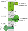
Fumarate respiration with NADH-quinone oxidoreductase (NuoA-N) and hydrogenase 2 (HybCOAB) as the primary dehydrogenases. The scheme gives the topology of the enzymes, including the sites for H+ release and consumption. For NADH-quinone reduction, a translocation of 4 H+/2 e is suggested. The reaction of fumarate reductase is not electrogenic (H+/2 e ratio of 0), whereas hydrogenase 2 is a redox-loop enzyme with a H+/2 e ratio of 2. More details on fumarate respiration, see Unden et al. (60) and Tomasiek et al. (91).
Figure 5
Figure 5
Anaerobic utilization of noncommon C4-dicarboxylates (L-tartrate, D-tartrate, D-malate) and relation to fumarate respiration. DcuB, C4-dicarboxylate/succinate antiporter; TtdT, L-tartrate/succinate antiporter; TtdAB, tartrate dehydratase; Mdh, malate dehydrogenase; FumB, (anaerobic) fumarase B; Frd, fumarate reductase; ET, electron transport; MKH2, menaquinol. Other details as described in Fig. 3 and in the text.
Figure 6
Figure 6
Stereoisomers of hydroxylated C4-dicarboxylates. The figure gives stereoisomers of C4-dicarboxylates carrying hydroxyl groups at C2, or at C2 and C3. Malate is found as L-malate (2S configuration) and D-malate (2R configuration), but only the L-isomer is of natural origin. Tartrate is represented by three stereoisomers (L-, D- and meso-tartrate). L-tartrate (2R, 3R configuration) is present in many plants, whereas D-tartrate (2S, 3S) is rare in nature and meso-tartrate not of natural origin. Fumarate and maleate are isomers of butenedioate. Maleate (cis-butenedioate) is chemically produced, whereas fumarate (trans-butenedioate) is a common intermediate of living cells.
Figure 7
Figure 7
Pathway, genes, and transcriptional regulation of the genes for citrate fermentation by citrate via the CitA-CitB and the DcuS-DcuB two-component systems. (A) Synthesis of the citrate fermentation specific enzymes and transporters (CitT, citrate/succinate antiporter; CL, citrate lyase)is induced by CitA-CitB and citrate (blue labeling). Synthesis of the fumarate respiration pathway (FrdABCD [or Frd], FumB, presented in green) is induced by the citrate response of DcuS-DcuR (using the side-activity of DcuS for citrate). (B) This scheme, for comparison, gives the fumarate respiratory system (FrdABCD, Frd) and fumarate/succinate antiporter (DcuB) that are induced by DcuS-DcuR in response to fumarate (or C4-dicarboxylates). The enzymes and the carrier shown in blue are unique for citrate fermentation; the enzymes shown in green are used both in citrate fermentation and fumarate respiration. Cit, citrate; Fum, fumarate; Mal, malate; OAA, oxaloacetate; Succ, succinate. Modified from reference .
Figure 8
Figure 8
Succinate production from endogenous fumarate (glucose fermentation). The scheme shows the major intermediates for the formation and excretion of succinate from PEP, and of formate, acetate, and ethanol formation (mixed acid fermentation) during glucose fermentation. Residual activities of the repressed and interrupted tricarboxylic acid (TCA) cycle are shaded with broken lines. AcCoA, acetyl-CoA; DcuC, succinate export carrier; FumB, (anaerobic) fumarase; FumC, (aerobic) fumarase; Frd, fumarate reductase; Mdh, NADH-dependent cytosolic malate dehydrogenase; OAA, oxaloacetate; PTS, PEP-dependent phosphotransferase uptake for glucose; PFL, pyruvate formate lyase; Ppc, PEP carboxylase; Pyr, pyruvate; Sdh, succinate dehydrogenase SdhABCD.
Figure 9
Figure 9
Succinate production pathways by engineered succinate production strains of E. coli. The scheme summarizes reactions engineered in E. coli to improve succinate production. Dotted arrows, either nonfunctional or decreased steps; Bold solid arrows, the primary route for carbon flow; Δ, gene deletion; +, overproduced or transformed genes; red letters, target genes; Ac-CoA, acetyl-CoA; Ace, acetate; Cit, citrate; DcuB, fumarate-succinate antiporter; DcuC, succinate export carrier; EMP, Embden-Meyerhof-Parnas pathway; EtOH, ethanol; For, formate; Fum, fumarate; GalP, galatose permease; G6P, glucose 6-phosphate; Isoc, isocitrate; Lac, lactate; Mal, malate; OAA, oxaloacetate; PEP, phosphoenolpyruvate; PTS, PEP-dependent phosphotransferase uptake for glucose; Pyr, pyruvate; Succ, succinate;. Genes: aceA, isocitrate lyase; aceB, malate synthase; ackA, acetate kinase; adhE, alcohol dehydrogenase; fumB, anaerobic fumarase; frdABCD, fumarate reductase; glk, glucokinase; iclR, isocitrate lyase regulator; ldh, lactate dehydrogenase; mdh, NADH-dependent malate dehydrogenase; pflB, pyruvate formate lyase; pk, pyruvate kinase; ppc, PEP carboxylase; pps, PEP synthethase; pta, phosphate acetyl transferase.
Figure 10
Figure 10
Phylogenetic tree of C4-dicarboxylate carriers of E. coli (DctA, DauA, DcuAB, DcuC, TtdT, CitT, and TRAP, but without SatP transporters) as derived from protein sequences. The distances represent the differences in identical amino acid residues of the carriers or the corresponding gene products. The amino acid sequences were aligned with Clustal Omega program (http://www.ebi.ac.uk/Tools/msa/clustalo/) and the tree was reconstructed by the neighbor-joining method MEGA 6.0 (213). Abbreviations of strains: As, Actinobacillus succinogenes; Bs, Bacillus subtilis; Cg, Corynebacterium glutamicum; Ec, E. coli; Ms, Mannheimia succinoproducens; Pa, Pseudomonas aeruginosa; Pf, Pseudomonas fluorescens; Ph, Pyrococcus horikoshii; Rl, Rhizobium leguminosarum; Rm, R. meliloti; Se, Salmonella enterica; Tk, Thermococus kodacarensis; Vc, Vibrio cholerae; Ws, Wolinella succinogens.
Figure 11
Figure 11
Carriers and transport modes for the exchange, uptake, and efflux of C4-dicarboxylates in E. coli under aerobic (A) and anaerobic growth conditions (B). (C) shows the transport modes for exchange, uptake, and efflux of C4-dicarboxylates that can be effected by the Dcu carriers (DcuA, DcuB, or DcuC). Uptake and efflux are electrogenic by the symport of 3 H+ with the C4-DC, whereas antiport is electroneutral (48). In aerobic growth (A), DctA is the major carrier for uptake at pH 7. DauA and potentially SatP replace DctA function at pH 5 and pH 6. The transporters catalyze electrogenic transport as presented in the figure. In anaerobic growth (B), during growth by fumarate respiration, DcuB is the major carrier and catalyzes an electroneutral fumarate/succinate antiport. DcuB can be supported or replaced by DcuA. During glucose fermentation, succinate efflux is effected by DcuC, which can be supported by DcuB, DcuA, and other unknown efflux carriers. During anaerobic growth on tartrate, tartrate-succinate antiport is catalyzed by carrier TtdT. The function of the dcuD gene product (DcuD) is unknown. References and details in the text. Modified from reference .
Figure 12
Figure 12
Domain structure and topology of the DcuS sensor kinase (A) and compaction (B) of the periplasmic citrate/C4dicarboxylate binding domains of CitA upon citrate binding or of DcuS upon L-malate binding, and (C) details of DctA/DcuS interaction. (A) DcuS is membrane-embedded by transmembrane helices 1 and 2 (TM1, TM2), and contains additionally the extracytoplasmic Per-Arndt-Sim domain PASP, a cytoplasmic PAS domain PASC, and a C-terminal HisKA/HATPase-type kinase. The monomers of the DcuS homodimer are presented in light and dark gray. In the dark gray monomer, the α-helical structure of TM2 and of the C-terminal helix α6 PASP is indicated. + and − indicate the periplasmic and cytoplasmic sides of the membrane. (B) Structure comparison of the periplasmic PASP domains of DcuS with L-malate (brown; #3BY8 [93]) and CitAKp without citrate (gold, #2V9A). Structures were superimposed using the software Chimera (214). More details are given in (93, 151, 196, 197). (C) For the DctA/DcuS complex, only monomers of the proteins are shown. DcuS is preferentially dimeric (195), whereas DctA is presumably a trimer (215). The C-terminal cytoplasmic helix 8b of DctA plays a central role in the interaction of DctA with DcuS (144). Helix 8b interacts with the PASC domain of DcuS and controls by the interaction the kinase activity of DcuS (see text for details).
Figure 13
Figure 13
Transmembrane signaling by DcuS: Control of the kinase activity by C4-dicarboxylates and the transporter proteins DctA (or DcuB). B and C represent the functional state of DcuS in the DctA/DcuS sensor complex (C4-dicarboxylate responsive DcuS). (A) shows DcuS without DctA (permanent active DcuS, constitutive ON). In the C4-dicarboxylate responsive state (B, C), binding of C4-dicarboxylates causes contraction of PASP with an uplift of α6 and of TM2 (red arrows) by one helical turn in TM2. The shift of TM2 is transmitted in the cytoplasm to PASC, causing relieved PASC dimerization and relief of kinase inhibition. DctA and PASC collaborate in silencing (or inhibiting) the activity of the kinase domain. PASC is only able to silence the kinase when properly positioned by DctA. DctA is therefore a cosilencer of PASc, and silencing of the kinase can be abolished artificially both by deletion of PASC or of DctA, or physiologically by the pulling of TM2 after C4-dicarboxylate binding at PASP. See text for references.
Figure 14
Figure 14
Coordinated regulation of common and noncommon C4-dicarboxylates metabolism mediated by DcuS-DcuR two-component system and LysR-type regulators TtdR and DmlR. The common C4-dicarboxylates and proteins/genes for general (central) C4-dicarboxylate metabolism are presented in gray, proteins and genes of the noncommon C4-dicarboxylate metabolism in orange. Dotted lines indicate that the type of regulation (direct or indirect) is not known. Arrow, induction; block, repression; common C4-dicarboxylates, gray square; D-malate, orange triangle; L-tartrate, orange circles.

References

    1. Boogerd FC, Boe L, Michelsen O, Jensen PR. 1998. atp Mutants of Escherichia coli fail to grow on succinate due to a transport deficiency. J Bacteriol 180:5855–5859. [PubMed] - PMC - PubMed
    1. Baker KE, Ditullio KP, Neuhard J, Kelln RA. 1996. Utilization of orotate as a pyrimidine source by Salmonella typhimurium and Escherichia coli requires the dicarboxylate transport protein encoded by dctA. J Bacteriol 178:7099–7105. [PubMed] - PMC - PubMed
    1. Kay WW, Kornberg HL. 1971. The uptake of C4-dicarboxylic acids by Escherichia coli. Eur J Biochem 18:274–281. 10.1111/j.1432-1033.1971.tb01240.x - DOI - PubMed
    1. Reed JL, Patel TR, Chen KH, Joyce AR, Applebee MK, Herring CD, Bui OT, Knight EM, Fong SS, Palsson BO. 2006. Systems approach to refining genome annotation. Proc Natl Acad Sci USA 103:17480–17484. [PubMed]10.1073/pnas.0603364103 - DOI - PMC - PubMed
    1. Janausch IG, Kim OB, Unden G. 2001. DctA- and Dcu-independent transport of succinate in Escherichia coli: contribution of diffusion and of alternative carriers. Arch Microbiol 176:224–230. [PubMed]10.1007/s002030100317 - DOI - PubMed

MeSH terms

LinkOut - more resources