. 2016 Jul 14;11(7):e0155888.
doi: 10.1371/journal.pone.0155888. eCollection 2016.

Circulating Extracellular RNA Markers of Liver Regeneration

Affiliations

Circulating Extracellular RNA Markers of Liver Regeneration

Irene K Yan et al. PLoS One. .

Abstract

Background and aims: Although a key determinant of hepatic recovery after injury is active liver regeneration, the ability to detect ongoing regeneration is lacking. The restoration of liver mass after hepatectomy involves systemic changes with coordinated changes in gene expression guiding regenerative responses, activation of progenitor cells, and proliferation of quiescent hepatocytes. We postulated that these responses involve intercellular communication involving extracellular RNA and that these could represent biomarkers of active regenerative responses.

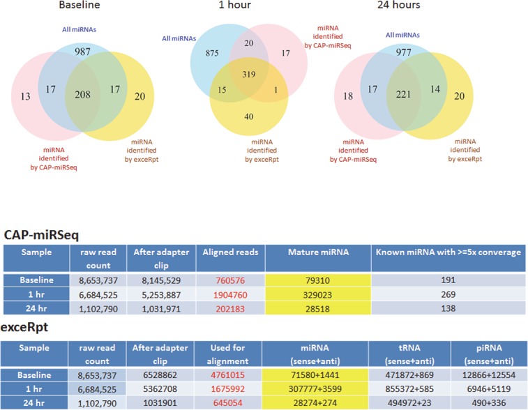
Methods: RNA sequencing was performed to identify temporal changes in serum extracellular non-coding RNA after partial hepatectomy in C57BL/6 male mice. Tissue expression of selected RNA was performed by microarray analysis and validated using qRT-PCR. Digital PCR was used to detect and quantify serum expression of selected RNA.

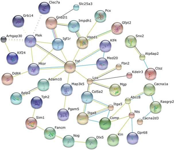
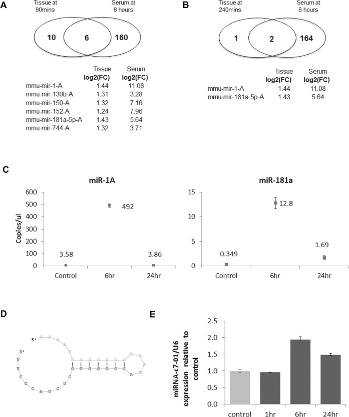
Results: A peak increase in extracellular RNA content occurred six hours after hepatectomy. RNA sequencing identified alterations in several small non-coding RNA including known and novel microRNAs, snoRNAs, tRNA, antisense and repeat elements after partial hepatectomy. Combinatorial effects and network analyses identified signal regulation, protein complex assembly, and signal transduction as the most common biological processes targeted by miRNA that altered. miR-1A and miR-181 were most significantly altered microRNA in both serum and in hepatic tissues, and their presence in serum was quantitated using digital PCR.

Conclusions: Extracellular RNA selectively enriched during acute regeneration can be detected within serum and represent biomarkers of ongoing liver regeneration in mice. The ability to detect ongoing active regeneration would improve the assessment of hepatic recovery from liver injury.


Conflict of interest statement

Competing Interests: The authors have declared that no competing interests exist.

Figures

Fig 1
Fig 1. Circulating extracellular RNA (exRNA) after partial hepatectomy.
(A). Serum was collected from mice at the indicated time points after partial hepatectomy, and exRNA was isolated. Total exRNA was quantitated using NanoDrop. An increase in circulating total exRNA was seen at six hours after partial hepatectomy. Bars represent average concentration of total exRNA yield from 4 separate samples. (B and C) RNA seq was performed on serum exRNA, and analysis of the serum exRNA was performed using the Maverix pipeline. Differentially-expressed extracellular non-coding RNA were determined across samples. (B) Non-coding RNA that are mapped include miRNA, tRNA, snoRNAs, antisense transcripts and repeat elements. (C). Bar chart representing the most highly differentially-expressed circulating EV non-coding RNA within selected classes six hours after partial hepatectomy. These differentially-expressed extracellular non-coding RNA are candidate biomarkers of a liver regenerative response.
Fig 2
Fig 2. Bioinformatics network analysis of miRNA target genes.
The target genes of the 52 miRNAs significantly altered in the circulation after partial hepatectomy were analyzed using the miRror program. Three hundred and five genes were predicted, and network analysis of these genes using String 9.05 indicated interactions amongst several identified targets.
Fig 3
Fig 3. Circulating extracellular RNA after partial hepatectomy.
(A and B) Comparison with tissue expression. miRNA expression was determined in liver tissues obtained at (A) 90 minutes and (B) 240 minutes after partial hepatectomy and were compared with miRNA detected in the serum six hours after partial hepatectomy. The Venn diagrams indicate miRNAs that had over 1.2 log-2 fold increase in both liver tissue and serum compared to baseline levels. The miRNA with the greatest increase in expression in both tissue and serum are indicated, with an increase in miR-1-A and miR 181a-5pA noted in hepatic tissue at both time points. (C) Quantitation by droplet digital PCR. A droplet digital PCR assay was used to analyze the expression of miR-1A, miR-181a-5p-A, and miR-222 in serum obtained at baseline, 6, and 24 hours after partial hepatectomy. exRNA was isolated from serum samples at each time point, treated with DNase, and reverse transcribed to obtain cDNA, and of which, 2ul was then emulsified into 1nL reaction droplets prior to target DNA amplification (40 cycles) performed using TaqMan miRNA assays. The fluorescence amplitude of each droplet was measured, and the target concentration (copies/ul) was calculated. No copies of miR-222 were detected. Data for other miRNAs represent merged data of mean quantitation from four technical replicates of one sample at each time point, with error bars showing the 95% confidence intervals after statistical analysis of positive and negative reactions based on poisson distribution. (D and E) Novel circulating extracellular miRNA. D. Putative miRNA with a genomic location mapped to chromosome 7 (pmiR-c7-01) identified by bioinformatics analysis of RNA sequencing data of circulating exRNA after partial hepatectomy. E. Serum expression of pmiR-c7-01 by qRT-PCR using custom- designed specific primers at indicated time points after partial hepatectomy.
Fig 4
Fig 4. Choice of analytic pipelines for identification of circulating exRNA.
(A and B). exRNA samples were obtained at baseline, and at 1 and 24 hours after partial hepatectomy. The number of aligned reads were sufficient for comparison for each sample using the Mayo CAP-miRSeq and Extracellular RNA Communication Consortium exceRpt analytical pipelines. A comparison was made with reference to all 1229 miRNAs that are similar between miRBase versions 11 and 12. (A) The Venn diagrams indicate the number of miRNA identified by each platform compared with all miRNAs. 16.9% of all miRNA were identified in the circulation at baseline by both platforms. This increased to 26.0% at 1 hour and was 18.0% at 24 hours. (B) The exceRpt platform also identified an increase in tRNA at 1 hour, as well as a dramatic reduction in piRNA at 1 and 24 hours compared with baseline.

References

    1. Michalopoulos GK. Principles of liver regeneration and growth homeostasis. Comprehensive Physiology. 2013;3(1):485–513. Epub 2013/05/31. 10.1002/cphy.c120014 . - DOI - PubMed
    1. Wei W, Dirsch O, McLean AL, Zafarnia S, Schwier M, Dahmen U. Rodent models and imaging techniques to study liver regeneration. European surgical research Europaische chirurgische Forschung Recherches chirurgicales europeennes. 2015;54(3–4):97–113. Epub 2014/11/18. 10.1159/000368573 . - DOI - PubMed
    1. Schug J, McKenna LB, Walton G, Hand N, Mukherjee S, Essuman K, et al. Dynamic recruitment of microRNAs to their mRNA targets in the regenerating liver. BMC genomics. 2013;14:264 Epub 2013/04/20. 10.1186/1471-2164-14-264 - DOI - PMC - PubMed
    1. Chiba M, Yasue H, Ohkohchi N. Gene expression profiling of sense and antisense transcripts in liver regeneration by microarray analysis. Biomedical reports. 2013;1(3):383–8. Epub 2014/03/22. 10.3892/br.2013.80 - DOI - PMC - PubMed
    1. Song G, Sharma AD, Roll GR, Ng R, Lee AY, Blelloch RH, et al. MicroRNAs control hepatocyte proliferation during liver regeneration. Hepatology. 2010;51(5):1735–43. Epub 2010/05/01. 10.1002/hep.23547 - DOI - PMC - PubMed