. 2017 Feb;139(2):462-471.e14.
doi: 10.1016/j.jaci.2016.04.037. Epub 2016 Jun 11.

IL-17A enhances IL-13 activity by enhancing IL-13-induced signal transducer and activator of transcription 6 activation

Affiliations

IL-17A enhances IL-13 activity by enhancing IL-13-induced signal transducer and activator of transcription 6 activation

Sara L Hall et al. J Allergy Clin Immunol. 2017 Feb.

Abstract

Background: Increased IL-17A production has been associated with more severe asthma; however, the mechanisms whereby IL-17A can contribute to IL-13-driven pathology in asthmatic patients remain unclear.

Objective: We sought to gain mechanistic insight into how IL-17A can influence IL-13-driven responses.

Methods: The effect of IL-17A on IL-13-induced airway hyperresponsiveness, gene expression, mucus hypersecretion, and airway inflammation was assessed by using in vivo models of IL-13-induced lung pathology and in vitro culture of murine fibroblast cell lines and primary fibroblasts and human epithelial cell lines or primary human epithelial cells exposed to IL-13, IL-17A, or both.

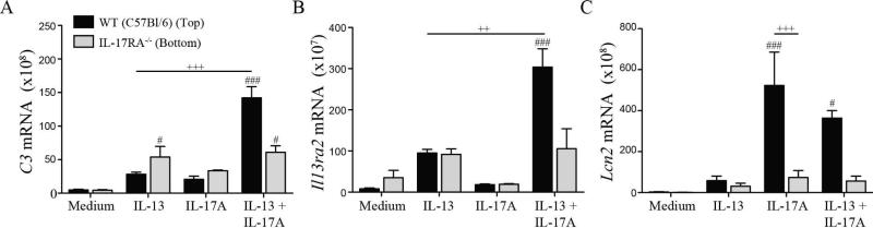
Results: Compared with mice given intratracheal IL-13 alone, those exposed to IL-13 and IL-17A had augmented airway hyperresponsiveness, mucus production, airway inflammation, and IL-13-induced gene expression. In vitro, IL-17A enhanced IL-13-induced gene expression in asthma-relevant murine and human cells. In contrast to the exacerbating influence of IL-17A on IL-13-induced responses, coexposure to IL-13 inhibited IL-17A-driven antimicrobial gene expression in vivo and in vitro. Mechanistically, in both primary human and murine cells, the IL-17A-driven increase in IL-13-induced gene expression was associated with enhanced IL-13-driven signal transducer and activator of transcription 6 activation.

Conclusions: Our data suggest that IL-17A contributes to asthma pathophysiology by increasing the capacity of IL-13 to activate intracellular signaling pathways, such as signal transducer and activator of transcription 6. These data represent the first mechanistic explanation of how IL-17A can directly contribute to the pathogenesis of IL-13-driven pathology.

Keywords: Asthma; IL-13; IL-17A; cytokines; signal transducer and activator of transcription 6; signal transduction.


Figures

Fig 1
Fig 1
Dose-dependent effects of IL-13 and IL-17A in vivo. AHR in A/J mice treated with IL-13 or IL-17A i.t. (A,B), total cell counts from BAL fluid of cytokine treated animals (C,D), frequency of macrophages, lymphocytes, neutrophils, and eosinophils in the BAL fluid (E,F), CLCA3 and αTubulin expression and densitometry in total lung homogenates (G), total lung expression of IL-13 (H) and IL-17A (I) induced transcripts following cytokine treatment. #, ##, and ### indicate P<0.05, <0.01, and < 0.001 vs PBS. +, ++, and +++ in indicate P<0.05, <0.01, and < 0.001 vs IL-13. Mean + SEM of n=4-9 mice per group (pooled over 3 independent experiments) are shown.
Fig 2
Fig 2
IL-13Rα2 is not required for IL-17A-mediated enhancement of IL-13-induced pathology. AHR in WT or IL-13Rα2−/− mice treated with IL-13 or IL-17A i.t. (A), total cell counts from BAL fluid of cytokine treated animals (B), frequency of macrophages, lymphocytes, neutrophils, and eosinophils in the BAL fluid (C,D), total lung expression of IL-13 (E) and IL-17A (F) induced transcripts following cytokine treatment. # and ## indicate P<0.05 and <0.01 vs PBS. + and +++ indicate P<0.05 and <0.001 vs IL-13 (A,B) or IL-17A (F). Mean + SEM of n=5-11 mice per group (pooled over 3 independent experiments) are shown.
Fig 3
Fig 3
Reciprocal co-regulation of IL-13- and IL-17A-induced genes occurs in vitro. Real-time PCR analysis of C3 (A), Il13ra2 (B), Cebpd (C), and Lcn2 (D) expression in NIH/3T3s stimulated with IL-13, IL-17A, or both cytokines over 24 hours. ## and ### indicate P<0.01 and <0.001 vs Medium. +, ++, and +++ in indicate P<0.05, <0.01, and < 0.001 vs IL-13 (A, B) or IL-17A (C). Mean + SEM of 4-8 replicates per condition shown. Results show 1 of 3 independent experiments.
Fig 4
Fig 4
IL-17A directly signals on IL-13-responsive cells to enhance gene expression. Real-time PCR analysis of C3 (A), Il13ra2 (B), and Lcn2 (C) expression in co-cultured WT or IL-17RA−/− lung fibroblasts stimulated with IL-13, IL-17A, or both cytokines for 24 hours. # and ### indicate P<0.05 and <0.001 vs Medium. ++ and +++ indicate P<0.01 and < 0.001 vs IL-13 (A, B) or IL-17A (C). Mean + SEM of 3 replicates per condition shown. Results show 1 of 3 independent experiments.
Fig 5
Fig 5
IL-17A does not stabilize IL-13-induced transcripts. Percentage mRNA remaining of IL-13 induced C3 (A) and Il13ra2 (B) in NIH/3T3s stimulated with IL-13 or IL-13 + IL-17A for 16 hours and incubated with ActD. Real-time PCR analysis of transcript levels was evaluated over 24 hours and t½ was calculated. Mean + SEM of 4-8 replicates per condition shown. Results show 1 of 3 independent experiments.
Fig 6
Fig 6
IL-17A directly enhances IL-13-driven pSTAT6. pSTAT6/STAT6 levels in NIH/3T3 cells pretreated with DMSO (A) or cycloheximide (B) then stimulated with cytokines for 5 minutes, (C) densitometry of pSTAT6/STAT6 expression shown in (A,B), geometric mean fluorescence intensity of pSTAT6 by phosphoflow in NIH/3T3s stimulated with cytokines over 1 hour (D), pSTAT6 and STAT6 expression and associated densitometry in whole lung homogenates from cytokine-treated A/J mice (mean + SEM of n=7-10 mice per group, pooled from 3 independent experiments) (E), real-time PCR analysis of C3 and Il13ra2 (F) or Lcn2 (G) expression in WT and STAT6−/− lung fibroblasts stimulated with cytokines for 24 hours. #, ##, and ### indicate P<0.05, <0.01, and <0.001 vs PBS or Medium. +, ++, and +++ indicate P<0.05 , <0.01, and <0.001 vs IL-13. Cyclohexamide experiments display 1 of 3 independent experiments performed. Phosphoflow cytometry and real-time PCR results show mean + SEM of 4-6 replicates per condition shown. Results show 1 of 2 independent experiments.
Fig 7
Fig 7
Reciprocal co-regulation by IL-13 and IL-17A is conserved in human cells. Real-time PCR analysis of SERPINB4 or IL13RA2 (A) and LCN2 (B) expression in primary human NECs (n=7) stimulated with IL-13, IL-17A, or both cytokines for 24 hours. pSTAT6 and STAT6 levels and associated densitometric analysis in Caco-2s (C), A549s (D) stimulated with cytokines for 5 minutes or NECs (n=2) stimulated with cytokines for 15 minutes (E). Caco-2 and A549 immunoblots are representative of 2 and 3 independent experiments, respectively.

References

    1. Grunig G, Warnock M, Wakil AE, Venkayya R, Brombacher F, Rennick DM, et al. Requirement for IL-13 independently of IL-4 in experimental asthma. Science. 1998;282:2261–3. - PMC - PubMed
    1. Walter DM, McIntire JJ, Berry G, McKenzie AN, Donaldson DD, DeKruyff RH, et al. Critical role for IL-13 in the development of allergen-induced airway hyperreactivity. J Immunol. 2001;167:4668–75. - PubMed
    1. Wills-Karp M, Luyimbazi J, Xu X, Schofield B, Neben TY, Karp CL, et al. Interleukin-13: central mediator of allergic asthma. Science. 1998;282:2258–61. - PubMed
    1. Zhu Z, Homer RJ, Wang Z, Chen Q, Geba GP, Wang J, et al. Pulmonary expression of interleukin-13 causes inflammation, mucus hypersecretion, subepithelial fibrosis, physiologic abnormalities, and eotaxin production. J Clin Invest. 1999;103:779–88. - PMC - PubMed
    1. Kuperman DA, Huang X, Koth LL, Chang GH, Dolganov GM, Zhu Z, et al. Direct effects of interleukin-13 on epithelial cells cause airway hyperreactivity and mucus overproduction in asthma. Nat Med. 2002;8:885–9. - PubMed

MeSH terms