. 2016 Sep 2;291(36):18867-79.
doi: 10.1074/jbc.M116.739227. Epub 2016 Jul 14.

Assembling of the Mycobacterium tuberculosis Cell Wall Core

Affiliations

Assembling of the Mycobacterium tuberculosis Cell Wall Core

Anna E Grzegorzewicz et al. J Biol Chem. .

Abstract

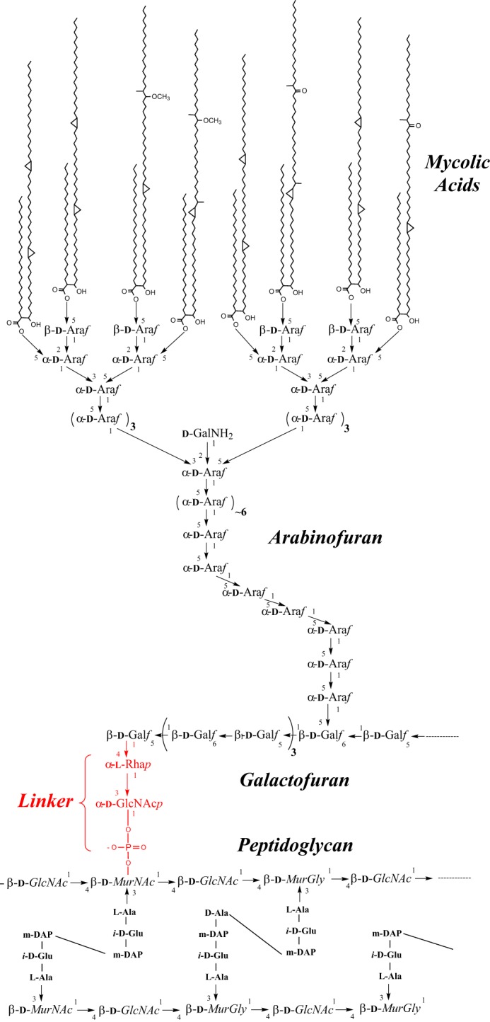
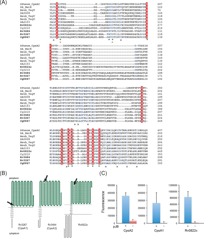
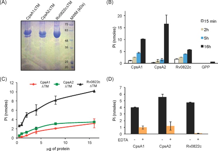
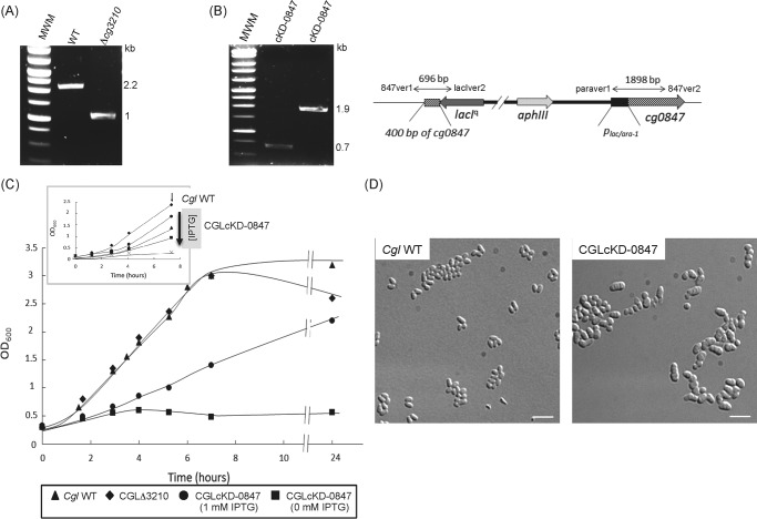
The unique cell wall of mycobacteria is essential to their viability and the target of many clinically used anti-tuberculosis drugs and inhibitors under development. Despite intensive efforts to identify the ligase(s) responsible for the covalent attachment of the two major heteropolysaccharides of the mycobacterial cell wall, arabinogalactan (AG) and peptidoglycan (PG), the enzyme or enzymes responsible have remained elusive. We here report on the identification of the two enzymes of Mycobacterium tuberculosis, CpsA1 (Rv3267) and CpsA2 (Rv3484), responsible for this function. CpsA1 and CpsA2 belong to the widespread LytR-Cps2A-Psr (LCP) family of enzymes that has been shown to catalyze a variety of glycopolymer transfer reactions in Gram-positive bacteria, including the attachment of wall teichoic acids to PG. Although individual cpsA1 and cpsA2 knock-outs of M. tuberculosis were readily obtained, the combined inactivation of both genes appears to be lethal. In the closely related microorganism Corynebacterium glutamicum, the ortholog of cpsA1 is the only gene involved in this function, and its conditional knockdown leads to dramatic changes in the cell wall composition and morphology of the bacteria due to extensive shedding of cell wall material in the culture medium as a result of defective attachment of AG to PG. This work marks an important step in our understanding of the biogenesis of the unique cell envelope of mycobacteria and opens new opportunities for drug development.

Keywords: Mycobacterium tuberculosis; arabinogalactan; cell wall; cryo-electron microscopy; ligase; outer membrane; peptidoglycan; polysaccharide.


Figures

FIGURE 1.
FIGURE 1.
The M. tuberculosis cell wall core. PG from M. tuberculosis is composed of linear chains of N-acetyl-α-d-glucosamine and modified muramic acid substituted with peptide side chains that are heavily cross-linked (70–80%), providing added structural integrity to the bacterium. AG is attached to PG through a phosphodiester link to position 6 of some of the Mur residues. The specific linker unit ensuring its covalent attachment to PG is shown in red. One arabinan chain is shown here attached to the galactan domain. The characteristic Ara6 non-reducing termini of the arabinan domain of AG serve as the anchoring points for the mycolates. Only one arabinan chain is shown for clarity.
FIGURE 2.
FIGURE 2.
Sequence and transmembrane topology of M. tuberculosis LCP protein candidates. A, alignment of the LytR-CpsA-Psr domains of LCP proteins and LCP protein candidates from M. tuberculosis, C. glutamicum, S. aureus, B. subtilis, and Streptococcus pneumoniae using Clustal Omega. Amino acids that are invariant in the alignment are colored white with a red background; homologous residues are in blue. Green diamonds, charged residues that contact the pyrophosphate headgroup of bound octaprenyl pyrophosphate in the crystal structure of CpsA2 from S. pneumoniae; orange diamonds, residues that coordinate a magnesium ion; crosses, conserved hydrophobic residues involved in the binding of the polyisoprenoid chain. B, transmembrane topology of Rv3267 (CpsA1), Rv3484 (CpsA2) and Rv0822c. The models were generated using TOPO2. C, topology of the C-terminal LCP domains of CpsA1, CpsA2, and Rv0822c in M. smegmatis. The LCP domains of the cpsA1, cpsA2, and Rv0822c genes were fused in frame with gfp in pJB(−) (red bars) and pJB(+) (blue bars). The positions of the GFP fusions in each protein are indicated by arrows and orange stars in the models shown in B. Fluorescence intensities were normalized to the A600 of the cultures, and the results shown represent the means and S.D. values of fluorescence intensities determined on at least 3–5 independent M. smegmatis transformants for each pJB(−) and pJB(+) plasmid. The addition of a single transmembrane domain from glycophorin A between the C-terminal fusion point of the protein of interest and the GFP in pJB(+) allows membrane-associated proteins with extracellular C-terminal fusions to be converted to proteins with intracellular C-terminal fusions. The native topology is reported with the fusion junction lacking the glycophorin A single transmembrane domain in the pJB(−) plasmid. Because GFP fluoresces in the cytoplasm but not in the periplasm, a high fluorescence signal in the pJB(+) version and background fluorescence in the pJB(−) version, as is the case with all three proteins here, are indicative of the C-terminal fusion of the protein being periplasmic. Error bars, S.D.
FIGURE 3.
FIGURE 3.
Pyrophosphatase activity of the three LCP proteins of M. tuberculosis recombinantly expressed and purified from E. coli. A, Coomassie Blue-stained SDS-polyacrylamide gel showing the purified CpsA1, CpsA2, and Rv0822c proteins devoid of transmembrane domain (CpsA1ΔTM, CpsA2ΔTM, and Rv0822cΔTM) upon affinity chromatography. The expected size of CpsA1ΔTM is 47.7 kDa, that of CpsA2ΔTM is 51.5 kDa, and that of Rv0822cΔTM is 52.4 kDa. Some degradation was consistently seen with CpsA1ΔTM and CpsA2ΔTM. The bottom bands have been confirmed to be proteolytic truncations of the target enzyme. B–D, pyrophosphatase assays. This assay monitors spectrophotometrically the hydrolysis of the pyrophosphate phosphoanhydride bond of GPP releasing Pi. B, the activity of the three purified LCP proteins is time-dependent. Reaction mixtures contained 5 μg (Rv0822cΔTM) or 10 μg (CpsA1ΔTM and CpsA2ΔTM) of recombinant proteins and were incubated for 15 min, 2 h, 5 h, and 16 h. C, protein concentration dependence. Reaction mixtures contained 0.5–16 μg of recombinant proteins and were incubated for 16 h. D, reaction mixtures contained 5 μg of recombinant protein and were incubated for 16 h in the absence (black bars) or presence (orange bars) of 10 mm EDTA. All assays were performed at least twice on independent recombinant protein preparations. Shown are the averages and S.D. values (error bars) of enzyme activities measured in duplicate in one representative experiment.
FIGURE 4.
FIGURE 4.
Inactivation of cg0847 and cg3210 in C. glutamicum. A, evidence for allelic replacement at the cg3210 locus of C. glutamicum. The deletion of the entire cg3210 ORF results in the replacement of the WT 2,157-bp amplification signal by a 1,033-bp fragment in the knock-out mutant. B, schematic representation of the chromosomal region of the cg0847 knockdown mutant (CGLcKD-0847) after integration of the pZEΔ847 plasmid into cg0847. Evidence for the correct insertion of the plasmid by PCR analysis using the two sets of primers, 847ver1/lacIver2 (resulting amplicon: 696 bp) and paraver1/847ver2 (resulting amplicon: 1,898 bp). C, growth characteristics of C. glutamicum WT, CGLΔ3210, and CGLcKD-0847. The strains were grown in LB broth at 30 °C with shaking. Inset, growth of CGLcKD-0847 in the presence of 0 (crosses), 12.5 μm (squares), 25 μm (triangles), or 1 mm (circles) IPTG; C. glutamicum WT (diamonds). The arrow indicates the time point at which samples were removed for the cell wall analyses described in this study. D, optical micrographs of C. glutamicum WT and CGLcKD-0847 cells cultivated overnight in BHI medium. One droplet of culture was absorbed onto a microscope slide coated with 1% agarose and visualized using a DMIRE2 optical microscope (Leica) equipped with a CCD camera (CoolSNAP HQ2, Roper Scientific). Scale bar, 5 μm.
FIGURE 5.
FIGURE 5.
Cryo-TEM of WT C. glutamicum (A) and the conditional knockdown strain CGLcKD-0847 (B). CGLcKD-0847 was grown in the presence of 25 μm IPTG. Black arrowheads, black arrows, and white arrows, plasma membrane, cell wall, and outer membrane, respectively. White arrowheads, detached membrane fragments in the conditional knockdown. Scale bar, 300 nm.
FIGURE 6.
FIGURE 6.
Analysis of the material released by CGLcKD-0847 in the culture medium. A, pellets obtained after ultracentrifugation of the culture supernatants from C. glutamicum WT or CGLcKD-0847 grown in the presence of 0, 0.025, or 1 mm IPTG. B, cryo-TEM of the material recovered upon ultracentrifugation of the culture supernatant of a C. glutamicum WT culture. Although there was no clearly visible pellet in this case, some material could be still recovered at the bottom of the tube. C, cryo-TEM of the material contained in the pellet shown in A for CGLcKD-0847. Vesicles seen in both WT and CGLcKD-0847 cultures are indicated by black arrows in B and C, and outer membrane fragments are indicated by white arrows in C. Scale bar, 100 nm. D, TLC analysis of total lipids extracted from whole C. glutamicum WT cells grown to stationary phase (lane 1) for comparison with lipids extracted from the pellet obtained in A (lane 2). TLC plates were developed in the solvent system CHCl3/CH3OH/H2O (65/25/4, by volume) and revealed by immersion in 10% H2SO4 in ethanol and heating. PG, phosphatidylglycerol; CL, cardiolipin; PIM, phosphatidylinositol mannosides; TMCM, trehalose monocorynomycolates; TDCM, trehalose dicorynomycolates. E, SDS-PAGE showing proteins extracted from the cell wall from WT C. glutamicum and CGLcKD-0847 grown in the presence of 1 mm IPTG or 25 μm IPTG and the protein composition of the pellet shown in A. MWM, molecular weight marker. F, sugar composition of the pellet obtained after ultracentrifugation of culture supernatants from CGLcKD-0847. Individual monosaccharides from the ultracentrifugation pellets of 300-ml CGLcKD-0847 cultures grown in the presence of 0 μm, 25 μm, and 1 mm IPTG were analyzed as their alditol acetate derivatives and their quantities (in μg) standardized to the A600 of the cultures. Although mannose was also detected, the precise quantification of this sugar was hampered by the large quantities of residual Glc, presumably coming from the culture medium.
FIGURE 7.
FIGURE 7.
Analysis of the linker regions from C. glutamicum WT, the cg0847 knockdown strain, and the cg3210 knock-out mutant. Shown are the ratios of MurNAc to MurNAc-6P as determined by LC/MS in cell wall samples prepared from C. glutamicum WT and CGLcKD-0847 grown in LB medium containing 0–1 mm IPTG (see Fig. 4C, inset) (A) and C. glutamicum WT and CGLΔ3210 grown to mid-log phase (A600 = 1.6–1.7) in LB medium (B).
FIGURE 8.
FIGURE 8.
Growth characteristics and cording properties of cpsA1 and cpsA2 knock-out mutants of M. tuberculosis H37Rv mc26206. A, evidence for allelic replacement at the cpsA1 and cpsA2 loci of M. tuberculosis H37Rv mc26206. The WT 2,175-bp amplicon is replaced by a 4,241-bp PCR fragment in the cpsA1 mutant due to the replacement of 158 bp of the cpsA1 ORF flanked between two AfeI restriction sites with a 2-kb streptomycin resistance cassette. The replacement of the entire cpsA2 gene by a 1.2-kb kanamycin resistance cassette results in the replacement of the WT 3,734-bp amplification signal by a 3,420-bp fragment in the mutant. B, growth characteristics of the cpsA1 and cpsA2 mutants of M. tuberculosis H37Rv mc26206 WT, the WT parent strain, and the complemented cpsA1 mutant, M. tuberculosis H37RvΔcpsA1/pMVGH1-cpsA1. The strains were grown in 7H9-ADC-Tween 80 broth at 37 °C with shaking. C, cording properties of the cpsA1 and cpsA2 mutants of M. tuberculosis H37Rv mc26206. Shown are Ziehl-Neelsen smears of 37 °C 7H9-ADC-tyloxapol cultures. Scale bar, 50.2 μm.

References

    1. Barry C. E., Crick D. C., and McNeil M. R. (2007) Targeting the formation of the cell wall core of Mycobacterium tuberculosis. Infect. Disord. Drug Targets 7, 182–202 - PMC - PubMed
    1. Jackson M., McNeil M. R., and Brennan P. J. (2013) Progress in targeting cell envelope biogenesis in Mycobacterium tuberculosis. Fut. Microbiol. 8, 855–875 - PMC - PubMed
    1. Daffé M., and Draper P. (1998) The envelope layers of mycobacteria with reference to their pathogenicity. Adv. Microb. Physiol. 39, 131–203 - PubMed
    1. Hoffmann C., Leis A., Niederweis M., Plitzko J. M., and Engelhardt H. (2008) Disclosure of the mycobacterial outer membrane: cryo-electron tomography and vitreous sections reveal the lipid bilayer structure. Proc. Natl. Acad. Sci. U.S.A. 105, 3963–3967 - PMC - PubMed
    1. Zuber B., Chami M., Houssin C., Dubochet J., Griffiths G., and Daffé M. (2008) Direct visualization of the outer membrane of mycobacteria and corynebacteria in their native state. J. Bacteriol. 190, 5672–5680 - PMC - PubMed

Publication types

MeSH terms

LinkOut - more resources