

Mucoepidermoid carcinoma arising in Warthin's tumor of the parotid gland: Clinicopathological characteristics and immunophenotypes
- PMID: 27417276
- PMCID: PMC4945913
- DOI: 10.1038/srep30149
Mucoepidermoid carcinoma arising in Warthin's tumor of the parotid gland: Clinicopathological characteristics and immunophenotypes
Abstract
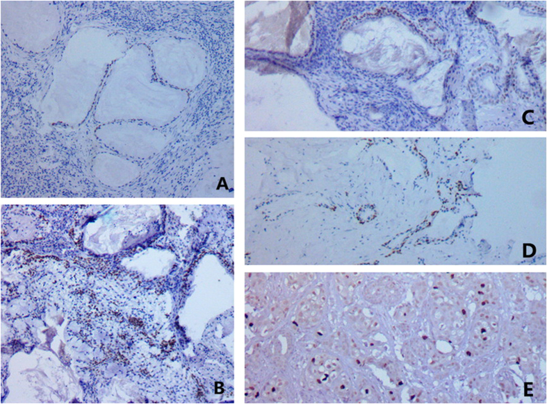
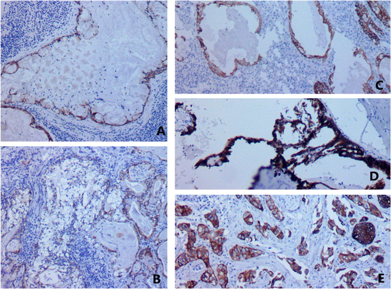
Mucoepidermoid carcinoma (MEC), an extremely rare tumor, arises from the epithelial component of preexisting parotid Warthin tumors (WT). Among the 309 cases of surgically resected WTs in Chinese PLA General Hospital and Beijing Shijitan Hospital of Capital Medical University, 5 cases (1.6%) fulfilled the criteria for MECs transformed from WTs. Clinicopathological characteristics of MECs was demonstrated in order to avoid misdiagnosis of this rare type of tumor. All the 5 patients, 3 males and 2 females, presented painless masses in the parotid gland. MECs were located inside or at the edge of WTs, with an obvious transitional zone between WT and MEC. Basal cells of WTs and epidermoid cells of MECs were strongly positive for cytokeratin CK5/6, CK34βE12, and P63; whereas negative for CK7, CK20, and CEA. Mucous cells of MECs were positive for CK7, CEA, as well as periodic acid-Schiff (PAS), whereas negative for CK5/6, CK34βE12, CK20, and P63. MECs patients were followed up for 25-69 months after surgery, presenting no evidence of recurrence or metastasis. Collectively, MECs arising from WT is very rare. The pathological diagnosis was based on histological morphology, especially the transitional zone between WT and MEC.
Figures


Similar articles
-
Mucoepidermoid carcinoma arising in Warthin's tumour of the parotid gland: report of two cases with histopathological, ultrastructural and immunohistochemical studies.Histopathology. 1998 Oct;33(4):379-86. doi: 10.1046/j.1365-2559.1998.00502.x. Histopathology. 1998. PMID: 9822930
-
Mucoepidermoid carcinoma arising in Warthin's tumor of the parotid gland.Pathol Int. 2002 Oct;52(10):653-6. doi: 10.1046/j.1440-1827.2002.01408.x. Pathol Int. 2002. PMID: 12445138
-
Mucoepidermoid carcinoma arising in Warthin's tumor: a case report.Indian J Pathol Microbiol. 2007 Apr;50(2):331-3. Indian J Pathol Microbiol. 2007. PMID: 17883061
-
Mucoepidermoid carcinoma involving Warthin tumor. A report of five cases and review of the literature.Am J Clin Pathol. 2000 Oct;114(4):564-70. doi: 10.1309/GUT1-F58A-V4WV-0D8P. Am J Clin Pathol. 2000. PMID: 11026102 Review.
-
Synchronous bilateral mucoepidermoid carcinoma of the parotid gland.J Laryngol Otol. 2003 May;117(5):419-21. doi: 10.1258/002221503321626537. J Laryngol Otol. 2003. PMID: 12803799 Review.
Cited by
-
Case report: The diagnostic pitfall of Warthin-like mucoepidermoid carcinoma.Front Oncol. 2024 Jun 20;14:1391616. doi: 10.3389/fonc.2024.1391616. eCollection 2024. Front Oncol. 2024. PMID: 38988706 Free PMC article.
-
Analysis of the Clinical Relevance of Histological Classification of Benign Epithelial Salivary Gland Tumours.Adv Ther. 2019 Aug;36(8):1950-1974. doi: 10.1007/s12325-019-01007-3. Epub 2019 Jun 17. Adv Ther. 2019. PMID: 31209701 Free PMC article. Review.
-
Warthin-like mucoepidermoid carcinoma of the parotid gland: a diagnostic and therapeutic dilemma.Autops Case Rep. 2019 Sep 30;9(4):e2019122. doi: 10.4322/acr.2019.122. eCollection 2019 Oct-Dec. Autops Case Rep. 2019. PMID: 31641663 Free PMC article.
-
Infarcted Warthin tumor with mucoepidermoid carcinoma-like metaplasia: a case report and review of the literature.J Med Case Rep. 2019 Jan 14;13(1):12. doi: 10.1186/s13256-018-1941-3. J Med Case Rep. 2019. PMID: 30636634 Free PMC article. Review.
-
MAML2 Rearrangements in Variant Forms of Mucoepidermoid Carcinoma: Ancillary Diagnostic Testing for the Ciliated and Warthin-like Variants.Am J Surg Pathol. 2018 Jan;42(1):130-136. doi: 10.1097/PAS.0000000000000932. Am J Surg Pathol. 2018. PMID: 28877061 Free PMC article.
References
-
- Bell D. & Luna M. A. Warthin adenocarcinoma: analysis of 2 cases of a distinct salivary neoplasm. Ann Diagn Pathol 13, 201–207 (2009). - PubMed
-
- Tian Z., Li L., Wang L., Hu Y. & Li J. Salivary gland neoplasms in oral and maxillofacial regions: a 23-year retrospective study of 6982 cases in an eastern Chinese population. Int J Oral Maxillofac Surg 39, 235–242 (2010). - PubMed
-
- Peter K. J. et al.. High risk for bilateral Warthin tumor in heavy smokers–review of 185 cases. Acta Otolaryngol 126, 1213–1217 (2006). - PubMed
-
- Gorai S. et al.. Malignant lymphoma arising from heterotopic Warthin’s tumor in the neck: case report and review of the literature. Tohoku J Exp Med 212, 199–205 (2007). - PubMed
MeSH terms
LinkOut - more resources
Full Text Sources
Other Literature Sources
Research Materials
