

Novel molecular insights into the critical role of sulfatide in myelin maintenance/function
- PMID: 27417284
- PMCID: PMC5037006
- DOI: 10.1111/jnc.13738
Novel molecular insights into the critical role of sulfatide in myelin maintenance/function
Abstract
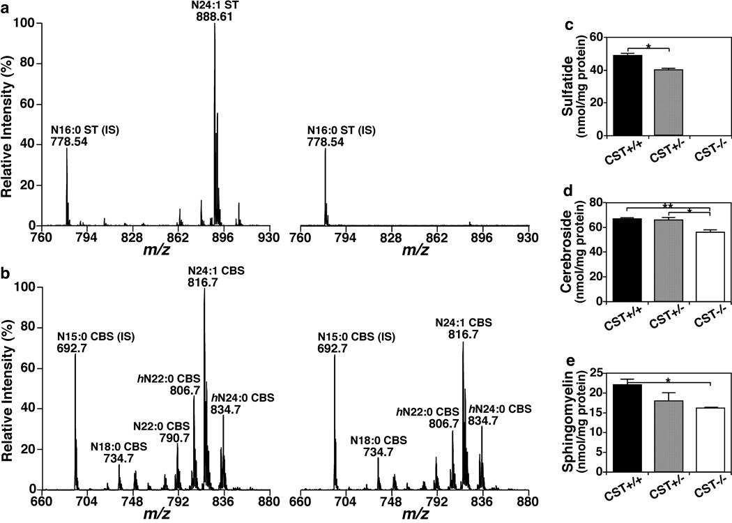
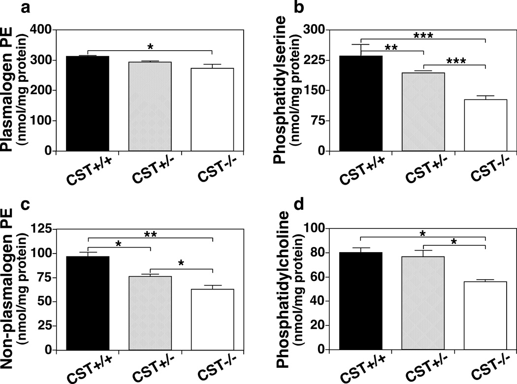
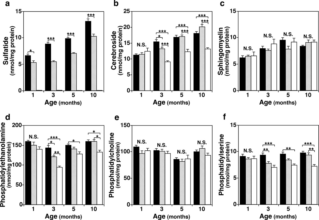
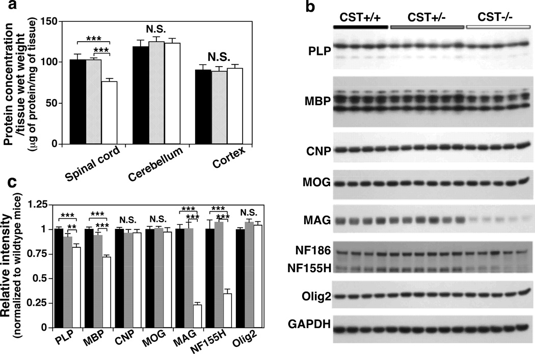
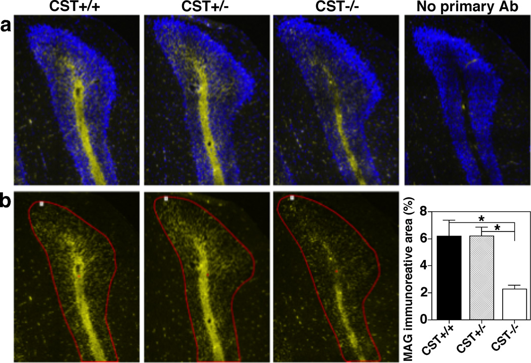
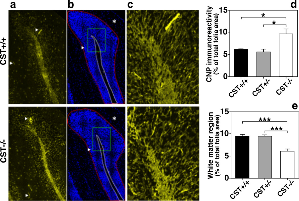
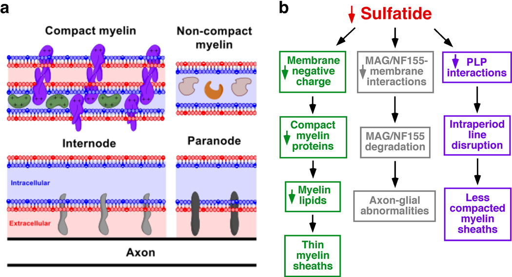
Cerebroside sulfotransferase (CST) catalyzes the production of sulfatide, a major class of myelin-specific lipids. CST knockout (CST(-/-) ) mice in which sulfatide is completely depleted are born healthy, but display myelin abnormalities and progressive tremors starting at 4-6 weeks of age. Although these phenotypes suggest that sulfatide plays a critical role in myelin maintenance/function, the underlying mechanisms remain largely unknown. We analyzed the major CNS myelin proteins and the major lipids enriched in the myelin in a spatiotemporal manner. We found a one-third reduction of the major compact myelin proteins (myelin basic protein, myelin basic protein, and proteolipid protein, PLP) and an equivalent post-developmental loss of myelin lipids, providing the molecular basis behind the thinner myelin sheaths. Our lipidomics data demonstrated that the observed global reduction of myelin lipid content was not because of an increase of lipid degradation but rather to the reduction of their synthesis by oligodendrocytes. We also showed that sulfatide depletion leads to region-specific effects on non-compact myelin, dramatically affecting the paranode (neurofascin 155) and the major inner tongue myelin protein (myelin-associated glycoprotein). Moreover, we demonstrated that sulfatide promotes the interaction between adjacent PLP extracellular domains, evidenced by a progressive decline of high molecular weight PLP complexes in CST(-/-) mice, providing an explanation at a molecular level regarding the uncompacted myelin sheaths. Finally, we proposed that the dramatic losses of neurofascin 155 and PLP interactions are responsible for the progressive tremors and eventual ataxia. In summary, we unraveled novel molecular insights into the critical role of sulfatide in myelin maintenance/function. Cerebroside sulfotransferase (CST) catalyzes the production of sulfatide, a major class of myelin-specific lipids. CST knockout (CST(-/-) ) mice in which sulfatide is completely depleted are born healthy, but display myelin abnormalities We show in our study that sulfatide depletion leads to losses of myelin proteins and lipids, and impairment of myelin functions, unraveling novel molecular insights into the critical role of sulfatide in myelin maintenance/function.
Keywords: cerebroside sulfotransferase; myelin lipidome; myelin-associated glycoprotein; proteolipid protein; shotgun lipidomics; sulfatide.
© 2016 International Society for Neurochemistry.
Conflict of interest statement
disclosure The authors have no conflict of interest to declare.
Figures
References
-
- Bansal R, Gard AL, Pfeiffer SE. Stimulation of oligodendrocyte differentiation in culture by growth in the presence of a monoclonal antibody to sulfated glycolipid. J Neurosci Res. 1988;21:260–267. - PubMed
-
- Bartsch S, Montag D, Schachner M, Bartsch U. Increased number of unmyelinated axons in optic nerves of adult mice deficient in the myelin-associated glycoprotein (MAG) Brain Res. 1997;762:231–234. - PubMed
-
- Benjamins JA, Morell P. Proteins of myelin and their metabolism. Neurochem Res. 1978;3:137–174. - PubMed
Publication types
MeSH terms
Substances
Grants and funding
LinkOut - more resources
Full Text Sources
Other Literature Sources
Molecular Biology Databases
Research Materials
