. 2016 Jul 15:7:12226.
doi: 10.1038/ncomms12226.

Identification of an ATP-controlled allosteric switch that controls actin filament nucleation by Arp2/3 complex

Affiliations

Identification of an ATP-controlled allosteric switch that controls actin filament nucleation by Arp2/3 complex

Max Rodnick-Smith et al. Nat Commun. .

Abstract

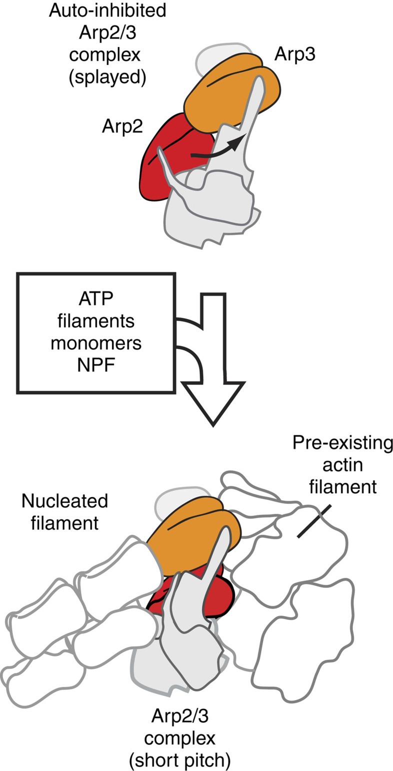
Nucleation of branched actin filaments by Arp2/3 complex is tightly regulated to control actin assembly in cells. Arp2/3 complex activation involves conformational changes brought about by ATP, Nucleation Promoting Factor (NPF) proteins, actin filaments and NPF-recruited actin monomers. To understand how these factors promote activation, we must first understand how the complex is held inactive in their absence. Here we demonstrate that the Arp3 C-terminal tail is a structural switch that prevents Arp2/3 complex from adopting an active conformation. The interaction between the tail and a hydrophobic groove in Arp3 blocks movement of Arp2 and Arp3 into an activated filament-like (short pitch) conformation. Our data indicate ATP binding destabilizes this interaction via an allosteric link between the Arp3 nucleotide cleft and the hydrophobic groove, thereby promoting the short-pitch conformation. Our results help explain how Arp2/3 complex is locked in an inactive state without activators and how autoinhibition is relieved.


Figures

Figure 1
Figure 1. Cartoon model of Arp2/3 complex activation.
Activation of Arp2/3 complex is thought to require movement of the actin-related subunits Arp2 and Arp3 from an inactive ‘splayed' conformation to an active conformation that mimics a filament-like short-pitch actin dimer. ATP, actin monomers, actin filaments and an NPF protein are typically required for activation.
Figure 2
Figure 2. The Arp3 C-terminal tail is a conserved structural feature required for autoinhibition of Arp2/3 complex.
(a) Ribbon diagram of Arp3 from crystal structure of Bos taurus Arp2/3 complex (1K8K) showing the position of the Arp3 C terminus (yellow). Subdomains 1–4 are numbered and the ATP-binding site (NBC) in Arp3 is indicated with cyan circle. The base and tip of the Arp3 C terminus are highlighted in pink and green, respectively. (b) Sequence alignment of the C terminus of Arp3 from diverse species. At, Arabidopsis thalania; Ce, Caenorhabditis elegans; Dm, Drosophila melanogaster; Hs, Homo sapiens; Dr, Danio rerio; Dd, Dictyostelium discoideum; Sc, Saccharomyces cerevisiae; Sp, Schizosaccharomyces pombe; act, rabbit skeletal muscle actin. Residues underlined and in red are deleted in arp3ΔC mutants. Cyan boxed residues are conserved in Arp3 sequences. Asterisks mark conserved hydrophobic residues that pin the C-terminal tail into the barbed end groove in some crystal structures. (c) Time course of polymerization of 3 μM 15 % pyrene-labelled actin showing constitutive activity of Arp3ΔC mutant compared with wild-type S. cerevisae (Sc)Arp2/3 complexes. (d) Maximum polymerization rate versus concentration of wild-type or Arp3ΔC S. cerevisiae complex for conditions described in c. (e) Time courses of pyrene actin polymerization as measured in b, but with S. pombe (Sp)Arp2/3 complexes. (f) Maximum polymerization rate versus concentration of wild-type or arp3ΔC S. pombe complexes for conditions described in e.
Figure 3
Figure 3. Deletion of the Arp3 C-terminal tail causes defects in endocytic actin patches and in the uptake of FM4-64.
(a) Spinning disk confocal fluorescence micrographs of wild type (WT) and mutant S. pombe cells expressing Fim1-GFP. Images are taken thorough the middle of the cells. Scale bar, 2 μm. (b) Average number of patches per cell (n≥50 cells). Error bars show s.e. (c) Kymographs of four actin patches for wild type and arp3ΔC strain. (d) Average patch lifetime for wild type and arp3ΔC strains. Error bars show s.e. (e) Quantification of Fim1-GFP intensity in WT and arp3ΔC S. pombe cells. Data show average intensity and s.d. from measurements of patches from wt (n=30 patches) and arp3ΔC (n=30) strains, respectively. The zero time point was taken to be the frame of maximum Fim1-GFP intensity during the lifetime of each patch. (f) FM4-64 endocytosis assay showing that arp3ΔC strain exhibits slowed dye uptake compared with the wild-type strain. Scale bar, 2 μm.
Figure 4
Figure 4. The Arp3 C terminus is required to prevent formation of the short-pitch conformation.
(a) Surface representation of BtArp2/3 complex (4JD2) showing the barbed end of Arp3, the barbed end groove and Arp3 C-terminal tail (yellow stick representation). Residues in Arp3 are coloured according to hydrophobicity, with polar residues orange and non-polar residues grey. Residues are labelled based on the S. cerevisiae Arp2/3 complex sequences. (b) Ribbon diagrams of Arp2 and Arp3 showing the position of engineered cysteines (Arp3(L155C), Arp2(R198C)) in the splayed and short-pitch conformations and the structure of the chemical crosslinker (BMOE) used in the short-pitch crosslinking assay. Structure 4JD2 was used to make both panels. In the right panel, the Oda et al. actin filament structure was used to move Arp2 into the short-pitch conformation. The solvent accessible crosslinking distance between engineered cysteines is 32.5 Å in the splayed conformation, and ranges from ∼8 to 11.3 Å in different models of the short-pitch conformation (Supplementary Fig. 3). BE: barbed end; PE: pointed end. (c) Anti-Arp3 western blot of 1-min crosslinking reactions containing 1 μM wild type (WT) or Arp3ΔC ScArp2/3 complexes in 200 μM ATP, 1 mM MgCl2, 50 mM KCl, 10 mM Imidazole pH 7.0, and 1 mM EGTA. Both complexes harbour the dual-engineered cysteine residues. The Arp3ΔC complex also contains the C426A mutation to eliminate the non-short-pitch crosslinking product (Fig. 7). (d) Quantification of reaction described in c. Error bars show s.e. for three reactions.
Figure 5
Figure 5. Destabilization of the splayed Arp2–Arp3 interface stimulates the short-pitch conformation and activates Arp2/3 complex.
(a) Ribbon diagram of Arp2 and Arp3 positioned in the splayed conformation (2P9K). Subdomains are numbered, with partially or fully disordered subdomains in Arp2 in parentheses. (b) Close up of the Arp2–Arp3 splayed interface, with key residues from Arp2 labelled based on S. cerevisiae sequence. (c) Coomassie stained gel of purified wild type and splayed interface mutants of ScArp2/3 complex. All budding yeast complexes in this panel and in subsequent figures harbour the engineered cysteine residues: (Arp2(R198C)/Arp3(L155C)). (d) Anti-Arp2/anti-Arp3 western blot of 1-min crosslinking reactions with 1 μM wild type or mutant ScArp2/3 complexes in 200 μM ATP, 1 mM MgCl2, 50 mM KCl, 10 mM Imidazole pH 7.0 and 1 mM EGTA. (e) Quantification of reaction described in d run in triplicate. Error bars show s.e. for three reactions. (f) Time course of 3 μM 15% pyrene actin polymerization with 25 nM wild type or splayed interface mutant Arp2/3 complexes as indicated. No NPF is present in these assays. (g) Maximum polymerization rate versus concentration of wild type or mutant ScArp2/3 complexes in pyrene actin polymerization assays as described in e.
Figure 6
Figure 6. The Arp3 C terminus is an allosteric molecular switch that locks the complex in the inactive (splayed) conformation.
(a) Cartoon depicting proposed conformational link between the ATP-binding cleft, the barbed end groove and the C terminus of Arp3. Dotted red lines indicate widths of nucleotide-binding cleft (NBC) and barbed end groove. The tip (T) and base (B) of the Arp3 C terminus are indicated. (b) Time course of crosslinking assays containing 1 μM wild-type ScArp2/3 complex with or without 100 μM Mg2+-ATP. (c) Quantification of 5-min short-pitch crosslinking for reactions containing 1 μM wild type or ATP-binding pocket mutation complexes with either 1 μM EDTA or 200 μM Mg2+-ATP. (d) Quantification of 1-min short-pitch crosslinking assays of wild type or splayed interface mutations with or without 200 μM Mg2+-ATP. Error bars in c and d show s.e. for three reactions.
Figure 7
Figure 7. Release of the tip of the Arp3 C terminus from the barbed end groove stimulates formation of the short-pitch conformation.
(a) Plot of average main chain B-factor versus residue number for the C-terminal residues in Arp3. Data taken from 10 different Bos taurus Arp2/3 complex crystal structures in different nucleotide-bound states (1K8K, 2P9L, 1U2V, 2P9N, 2P9U, 2P9P, 1TYQ, 2P9I, 2P9K and 2P9S; 14–16). Nucleotide cleft widths are classified based on Nolen and Pollard. B-factors were normalized so that each chain had the same average main chain B-factor. Residues missing in the electron density are omitted from the plot. Residue numbers in parenthesis are for ScArp2/3 complex. See Supplementary Fig. 6 for more detailed B-factor analysis. (b) Surface representation of the barbed end groove with C terminus of Arp3 bound (yellow stick representation, black labels) based off of structure 1K8K. Orange labels indicate key hydrophobic residues in the Arp3-barbed end groove. C426, a cysteine that becomes reactive upon deletion of the Arp3 C-terminal tail, is labelled in magenta. Residue labels are based on S. cerevisiae sequence. Black line indicates transition between the base and tip of the C-terminal tail. (c) Time courses of polymerization of 3 μM 15% pyrene actin in the presence of 10 nM wild type, Arp3ΔC, Arp3ΔC(C426A) or Arp3(L445D/F446D) ScArp2/3 complexes. The C426A mutation decreased activity slightly in the Arp3ΔC complex, but was still hyperactive compared with wild type. (d) Maximum polymerization rate versus complex concentrations for reactions described in c. (e) Time course of short-pitch crosslinking for reactions containing 1 μM wild type, Arp3ΔC or Arp3(L445D and F446D) ScArp2/3 complexes with or without 200 μM ATP. Error bars show s.e. for at least three reactions. (f) Two colour (anti-Arp2/anti-Arp3) western blot of crosslinking reactions containing 1 μM wild type or mutant ScArp2/3 complexes with or without 200 μM ATP as indicated. Reactions containing wild type and Arp3(L445D/F446D) complexes with or without ATP were run for 5 min. Reactions containing Arp3ΔC or Arp3ΔC(C426A) complexes were run for 1 min.

Similar articles

Cited by

References

    1. Goley E. D. & Welch M. D. The ARP2/3 complex: an actin nucleator comes of age. Nat. Rev. Mol. Cell Biol. 7, 713–726 (2006). - PubMed
    1. Rotty J. D., Wu C. & Bear J. E. New insights into the regulation and cellular functions of the ARP2/3 complex. Nat. Rev. Mol. Cell Biol. 14, 7–12 (2013). - PubMed
    1. Yi K. et al.. Dynamic maintenance of asymmetric meiotic spindle position through Arp2/3-complex-driven cytoplasmic streaming in mouse oocytes. Nat. Cell Biol. 13, 1252–1258 (2011). - PMC - PubMed
    1. Achard V. et al.. A ‘primer'-based mechanism underlies branched actin filament network formation and motility. Curr. Biol. 20, 423–428 (2010). - PubMed
    1. Marchand J. B., Kaiser D. A., Pollard T. D. & Higgs H. N. Interaction of WASP/Scar proteins with actin and vertebrate Arp2/3 complex. Nat. Cell Biol. 3, 76–82 (2001). - PubMed

Publication types

MeSH terms

Substances