. 2016 Jul 15:7:12116.
doi: 10.1038/ncomms12116.

Structural investigation of heteroyohimbine alkaloid synthesis reveals active site elements that control stereoselectivity

Affiliations

Structural investigation of heteroyohimbine alkaloid synthesis reveals active site elements that control stereoselectivity

Anna Stavrinides et al. Nat Commun. .

Abstract

Plants produce an enormous array of biologically active metabolites, often with stereochemical variations on the same molecular scaffold. These changes in stereochemistry dramatically impact biological activity. Notably, the stereoisomers of the heteroyohimbine alkaloids show diverse pharmacological activities. We reported a medium chain dehydrogenase/reductase (MDR) from Catharanthus roseus that catalyses formation of a heteroyohimbine isomer. Here we report the discovery of additional heteroyohimbine synthases (HYSs), one of which produces a mixture of diastereomers. The crystal structures for three HYSs have been solved, providing insight into the mechanism of reactivity and stereoselectivity, with mutation of one loop transforming product specificity. Localization and gene silencing experiments provide a basis for understanding the function of these enzymes in vivo. This work sets the stage to explore how MDRs evolved to generate structural and biological diversity in specialized plant metabolism and opens the possibility for metabolic engineering of new compounds based on this scaffold.


Figures
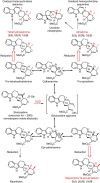
Figure 1
Figure 1. Heteroyohimbine alkaloid biosynthesis.
Heteroyohimbines with 3(S) stereochemistry derive from strictosidine aglycone. The three diastereomers found in Catharanthus roseus, are highlighted with red arrows. Alkaloids derived from heteroyohimbines are also shown.
Figure 2
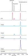
Figure 2. LC-MS analysis of active MDR candidates against strictosidine aglycone.
Cr033062 exhibited only trace activity. See Supplementary Fig. 1 for chromatograms of assays with inactive enzymes and negative controls.
Figure 3
Figure 3. Sequence alignment of Catharanthus roseus HYS enzymes and Populus tremuloides SAD
Numbering corresponds to HYS. Identical and similar amino acids are highlighted in red and yellow, respectively. Secondary structure elements of the HYS apo crystal structure are displayed. THAS1- and HYS-active site amino acids (Y56/53 and E59/56) are indicated by blue dots, and THAS2 active site amino acids (Y120 and D49) are indicated by green dots. Ligands for catalytic and structural zinc ions are highlighted by black and grey dots, respectively. The nuclear localization signal of (THAS1 and HYS) and loops 1 and 2, respectively, are indicated in red. A non-proline cis-peptide bond that is observed in THAS1 holo, in one subunit of THAS1 apo (Supplementary Fig. 5), in HYS apo, and not at all in THAS2 is indicated with an orange dot. The substrate-binding domain and the cofactor-binding domain are indicated by blue and purple bars, respectively.
Figure 4
Figure 4. Crystal structures of heteroyohimbine synthases THAS1, THAS2 and HYS.
(a) Sample of automatically derived experimentally phased electron density from THAS1 (at 1.12 Å resolution) superimposed on the final model showing the active site region with the NADP+ cofactor (green carbons) together with neighbouring residues (magnolia carbons) and water molecules (small red spheres). (b) THAS1 docked with cathenamine (pale blue carbons) with the protein shown in both cartoon (left) and space filling (right) modes. The NADP+ cofactor is shown with green carbons; loop 1 is in orange and loop 2 is in cyan. Zinc ions are displayed as magenta spheres. The active site is largely contained within a single subunit (magnolia surface), although the mouth of the channel leading to the active site is partially bounded by the second subunit of the biological dimer (grey surface) (c) Superposition of the apo structures of THAS1 (gold), THAS2 (pink) and HYS (blue). (d) Superposition of the holo (NADP+ containing) structures of THAS1 (gold) and THAS2 (pink), with the cofactor of THAS1 shown as van der Waals spheres for emphasis. For c and d, the structures were superposed onto the THAS1 structure, based on the upper subunit alone; only part of the lower subunit of the THAS1 structure is shown in grey for reference (see Supplementary Fig. 3 for images of the full THAS1 dimer). The insets emphasize the differing lengths of loop 2 between the various structures; the central portion of loop 2 in apo THAS2 was disordered.
Figure 5
Figure 5. Mechanistic hypothesis for heteroyohimbine synthases.
(a) Proposed mechanism for formation of the tetrahydroalstonine (S C20) diastereomer. (b) Proposed mechanism of formation of the ajmalicine (R C20) diastereomer that is observed in HYS, which contains a histidine residue near the active site.
Figure 6
Figure 6. Deuterium labelling of THAS1 and HYS products using pro-R-NADPD.
Comparison of selected regions of 1H-NMR spectra of labelled (a) tetrahydroalstonine, (b) ajmalicine, (c) mayumbine. The spectra indicate that C21 is labelled with deuterium in the pro-R position.
Figure 7
Figure 7. Product profiles of THAS1 and HYS loop swap mutants.
(a) Shown is the apo THAS1 structure (magnolia) with loops 1 and 2 highlighted in orange and cyan, respectively. For clarity, only the corresponding loops of the HYS apo structure are shown in yellow after superposition. Similarly, only the cofactor from the superposed holo THAS1 structure is shown for reference (green carbons). The side chains of important residues are also shown. (b) Representative LC-MS chromatograms of assays with THAS1 mutants in which loop 1, loop 2 or both have been swapped with the corresponding sequences from HYS. (c) Representative LC-MS chromatograms of assays with HYS mutants in which loop 1, loop 2 or both have been swapped with the corresponding sequences from THAS1.
Figure 8
Figure 8. THAS2 displays nucleocytosolic localization while HYS is preferentially targeted to the nucleus.
C. roseus cells were transiently co-transformed with plasmids expressing either THAS2–YFP (a), HYS–YFP (e) or YFP (i) and the plasmid encoding the nuclear-CFP marker(b,f,j). Co-localization of the fluorescence signals appears in yellow when merging the two individual (green/red) false colour images (c,g,k). Cell morphology is observed with differential interference contrast (DIC) (d,h,l). Scale bars, 10 μm.
Figure 9
Figure 9. THAS2 and HYS interact with SGD in the nucleus.
THAS2/SGD (a,i,q) and HYS/SGD (c,k,s) interactions were analysed by BiFC in C. roseus cells transiently transformed by distinct combinations of plasmids encoding fusions with the two split YFP fragments, as indicated on each fluorescence picture. THAS1/SGD (e,m,u) and 16OMT/SGD (g,o,w) interactions were studied to evaluate the specificity of THAS2/SGD and HYS/SGD interactions. Single BiFC assays showing interactions with SGD (upper row) and double BiFC assays highlighting both interactions with SGD and THAS2, HYS, THAS1, 16OMT self-interactions were conducted and observed 16 h (middle row) and 36 h (lower row) post-transformation. Cell morphology is observed with differential interference contrast (DIC) (b,d,f,h,j,l,n,p,r,t,v,x). Scale bars, 10 μm.

Similar articles

Cited by

References

    1. Shamma M. & Richey J. M. The stereochemistry of the heteroyohimbine alkaloids. J. Am. Chem. Soc. 85, 2507–2512 (1963).
    1. Allain H. & Bentue-Ferrer D. Clinical efficacy of almitrine-raubasine. An overview. Eur. Neurol. 39, 39–44 (1998). - PubMed
    1. Benzi G. Pharmacological features of an almitrine-raubasine combination. Activity at cerebral levels. Eur. Neurol. 39, 31–38 (1998). - PubMed
    1. Li S. et al.. Assessment of the therapeutic activity of a combination of almitrine and raubasine on functional rehabilitation following ischaemic stroke. Curr. Med. Res. Opin. 20, 409–415 (2004). - PubMed
    1. Roquebert J. & Demichel P. Inhibition of the a1- and a2-adrenoceptor-mediated pressor response in pithed rats by raubasine, tetrahydroalstonine and akuammigine. Eur. J. Pharmacol. 106, 203–205 (1984). - PubMed

Publication types

MeSH terms