Review
. 2016 Oct;100(4):687-698.
doi: 10.1189/jlb.3RU0316-151R. Epub 2016 Jul 14.

Endotoxemia-menace, marker, or mistake?

Affiliations
Review

Endotoxemia-menace, marker, or mistake?

Robert S Munford. J Leukoc Biol. 2016 Oct.

Abstract

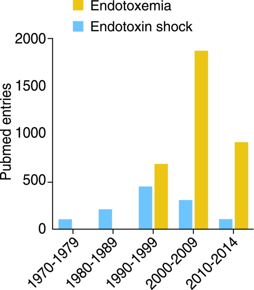
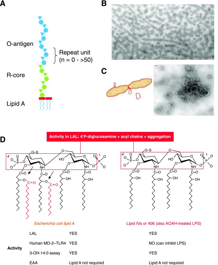
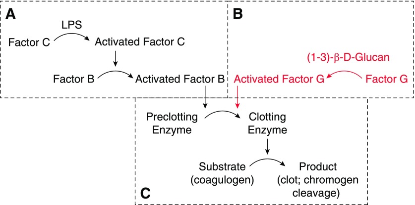
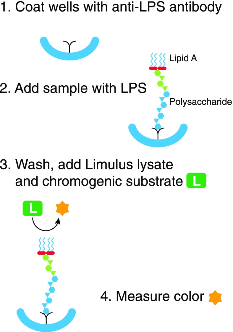
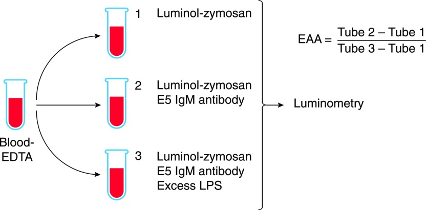
Endotoxemia is in its scientific ascendancy. Never has blood-borne, Gram-negative bacterial endotoxin (LPS) been invoked in the pathogenesis of so many diseases-not only as a trigger for septic shock, once its most cited role, but also as a contributor to atherosclerosis, obesity, chronic fatigue, metabolic syndrome, and many other conditions. Finding elevated plasma endotoxin levels has been essential supporting evidence for each of these links, yet the assays used to detect and quantitate endotoxin have important limitations. This article describes several assays for endotoxin in plasma, reviews what they do and do not measure, and discusses why LPS heterogeneity, LPS trafficking pathways, and host LPS inactivation mechanisms should be considered when interpreting endotoxin assay results.

Keywords: LPS; Limulus; endotoxin; lipopolysaccharide; translocation.


Figures

Figure 1.
Figure 1.. Publications with “endotoxemia” or “endotoxin shock” as keywords.
Data tabulated from PubMed entries with these Medical Subject Headings, by decade. Note that the columns for 2010–2014 include entries for only 5 yr. There were 345 entries for endotoxemia in 2015.
Figure 2.
Figure 2.. LPS structure, lipid A structure-activity relationships.
(A) Diagram showing the structure of a typical LPS, with lipid A, R-core oligosaccharide, and O-antigen moieties. (B) Salmonella typhimurium LPS stained with uranyl acetate, ×98,000. Note tubular and globular forms. Originally published in Shands and Chun [5], © American Society for Biochemistry and Molecular Biology. (C) Diagram showing release of outer membrane vesicles (blebs) by growing Escherichia coli. Some blebs appear at the time of septum closure during cell division. Negatively stained electron micrograph of an S. typhimurium culture supernatant showing blebs. Original bar (upper left), 100 nm. Originally published in Vesy et al. [43], © 2000, American Society for Microbiology. (D) Maximal lipid A sensing by human MD-2–TLR4 requires phosphates at both 1 and 4′ on the diglucosamine backbone, 4 glucosamine-linked acyl chains, and 2 secondary (acyloxyacyl) chains (red). Both E. coli lipid A and lipid IVa (also known as compound 406) are active in the LAL assay, which requires the 4′ phosphoryl diglucosamine backbone and aggregated acyl chains. In contrast, aggregation is not necessary for lipid A with an attached O-antigen to activate cells via MD-2–TLR4. Both lipid IVa and AOAH-treated LPS have a tetraacyl lipid A structure (right) and can inhibit LPS signaling, but AOAH-treated LPS is much less active than lipid IVa in the LAL. Note in both structures that the 4 3-OH-14:0 chains are all attached to the backbone; releasing them, using hot acid hydrolysis, has been used to discriminate LPS-linked and non-LPS 3-OH-14:0 in plasma (see text). S, Attachment site of polysaccharide chain (missing in lipid IVa but not in AOAH-treated LPS); arrows, sites of cleavage by AOAH.
Figure 3.
Figure 3.. The LAL assay.
The Limulus amebocyte contains a clotting enzyme cascade that can be initiated by (A) LPS binding factor C or (B) (1–3)-β-d-glucan binding factor G. Both pathways initiate cleavage of the proclotting enzyme to form the clotting enzyme (C). LPS may also be detected using recombinant factor C with its chromogenic substrate. A widely used clinical assay for β-glucan is based on the enzymes in B.
Figure 4.
Figure 4.. The “immunolimulus” assay.
Microtiter wells are first coated with an antibody to the target LPS (1). After washing, plasma is added. LPS in the plasma binds to the antibody (2). After another thorough wash, Limulus lysate and a chromogenic peptide substrate are added (3). The color produced is measured over time (4). The approach makes the LAL assay more specific for LPS.
Figure 5.
Figure 5.. The EAA.
Anticoagulated blood is incubated with an excess of LPS in tube 3 before E5 IgM antibody is added to tubes 2 and 3 and luminol-zymosan is added to all tubes. After incubation at 37°C with shaking, photons are counted using a luminometer. EAA is calculated using the formula shown. Graphic adapted from Matsumoto et al. [85].

References

    1. Suffredini A. F., Fromm R. E., Parker M. M., Brenner M., Kovacs J. A., Wesley R. A., Parrillo J. E. (1989) The cardiovascular response of normal humans to the administration of endotoxin. N. Engl. J. Med. 321, 280–287. - PubMed
    1. Marshall J. C. (2005) Lipopolysaccharide: an endotoxin or an exogenous hormone? Clin. Infect. Dis. 41 (Suppl 7), S470–S480. - PubMed
    1. Hurley J. C., Nowak P., Öhrmalm L., Gogos C., Armaganidis A., Giamarellos-Bourboulis E. J. (2015) Endotoxemia as a diagnostic tool for patients with suspected bacteremia caused by gram-negative organisms: a meta-analysis of 4 decades of studies. J. Clin. Microbiol. 53, 1183–1191. - PMC - PubMed
    1. Opal S. M., Scannon P. J., Vincent J. L., White M., Carroll S. F., Palardy J. E., Parejo N. A., Pribble J. P., Lemke J. H. (1999) Relationship between plasma levels of lipopolysaccharide (LPS) and LPS-binding protein in patients with severe sepsis and septic shock. J. Infect. Dis. 180, 1584–1589. - PubMed
    1. Shands J. W. Jr.,Chun P. W. (1980) The dispersion of gram-negative lipopolysaccharide by deoxycholate. Subunit molecular weight. J. Biol. Chem. 255, 1221–1226. - PubMed

Publication types

MeSH terms