Review
. 2016 Jul;11(7):919-39.
doi: 10.2217/fmb-2016-0044. Epub 2016 Jul 15.

Right on Q: genetics begin to unravel Coxiella burnetii host cell interactions

Affiliations
Review

Right on Q: genetics begin to unravel Coxiella burnetii host cell interactions

Charles L Larson et al. Future Microbiol. 2016 Jul.

Abstract

Invasion of macrophages and replication within an acidic and degradative phagolysosome-like vacuole are essential for disease pathogenesis by Coxiella burnetii, the bacterial agent of human Q fever. Previous experimental constraints imposed by the obligate intracellular nature of Coxiella limited knowledge of pathogen strategies that promote infection. Fortunately, new genetic tools facilitated by axenic culture now allow allelic exchange and transposon mutagenesis approaches for virulence gene discovery. Phenotypic screens have illuminated the critical importance of Coxiella's type 4B secretion system in host cell subversion and discovered genes encoding translocated effector proteins that manipulate critical infection events. Here, we highlight the cellular microbiology and genetics of Coxiella and how recent technical advances now make Coxiella a model organism to study macrophage parasitism.

Keywords: Coxiella burnetii; apoptosis; autophagy; effector protein; host cell invasion; macrophages; mutagenesis; phagolysosome; type 4B secretion; vacuole remodeling.


Conflict of interest statement

Financial & competing interests disclosure

This work was supported by the Intramural Research Program of the NIH, National Institute of Allergy and Infectious Diseases (RA Heinzen) and by grants from the Agence Nationale de la Recherche (ANR) (ANR-14-CE14-0012-01; Project AttaQ), ERA-NET Infect-ERA (ANR-13-IFEC-0003; Project EUGENPATH) and the ATIP-AVENIR programme (M Bonazzi). The authors have no other relevant affiliations or financial involvement with any organization or entity with a financial interest in or financial conflict with the subject matter or materials discussed in the manuscript apart from those disclosed.

No writing assistance was utilized in the production of this manuscript.

Figures

<b>Figure 1.</b>
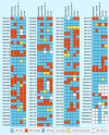
Figure 1.. Polymorphisms of Coxiella effector genes.
ORFs encoding validated Dot/Icm substrates were used to BLAST the genomes of the Nine Mile Phase I (NMI) RSA493, Henzerling RSA331, Dugway 5J108-111, G Q212 and K Q154 strains for homologous sequences. Effector ORFs were classified as intact (blue) if the length of the translated amino acid sequence was at least 90% of the longest representative amino acid sequence. Homologous ORFs with polymorphisms leading to protein sequence lengths less than 90% of the longest representative amino acid sequence were considered disrupted (red). In some cases, an intact effector gene was present but not annotated in GenBank (blue with slash), or completely missing (white). Amino acid sequences encoded by intact ORFs were further inspected for C-terminal polymorphisms, resulting largely from frame shift mutations, predicted to disrupt the translocation signal (yellow). Only 44 effector ORFs are conserved among the Coxiella strains in this analysis. The ORF identifiers CBU and CBUA correspond to chromosomal and plasmid genes, respectively, of the NMI RSA493 strain. The ORF identifiers CBUD and CBUDA correspond to chromosomal and plasmid genes, respectively, of the Dugway 5J108-111 strain. ORF: Open reading frame.
<b>Figure 2.</b>
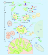
Figure 2.. Steps in Coxiella infection and Coxiella-containing vacuole formation and host cell processes potentially modulated by Coxiella effector proteins.
Coxiella internalization is facilitated by the rearrangement of the host actin cytoskeleton, mediated by the actin-binding proteins cortactin and mDia and requiring the activation of the small GTP-ases RhoA and ROCK. The Coxiella invasin OmpA mediates bacterial internalization by nonprofessional phagocytes, whereas internalization by macrophages requires interaction with cell surface αVβ3 integrins. Inside cells bacteria reside within CCVs that progressively acquire endosomal markers by heterotypic fusion with early and late endosomes and lysosomes (EE, LE and Ly, respectively). CCV acidification activates bacterial metabolism, which likely coincides with translocation of effectors (blue dots) by the Dot/Icm T4BSS (purple rods). This is required for vacuole expansion and for the recruitment of the autophagosomal and endocytic SNARE proteins syntaxin-17 and Vamp7, mediating the homotypic fusion of multiple CCVs into a single vacuole. CCV maturation requires Coxiella effectors to reroute several vesicle trafficking pathways that contribute membrane to the expanding CCV. The Coxiella effector CvpA interacts with AP2 on RE. The effector CvpB localizes at EE and CCVs, where it binds PI(3)P (red dots) and manipulates its metabolism to favor vacuole biogenesis and autophagy (Au)-mediated homotypic fusion of CCVs. Vacuole decoration by the small GTPase Rab1b suggests that other Coxiella effectors manipulate ER-to-Golgi traffic. Additional Coxiella effectors inhibit the intrinsic and extrinsic apoptotic pathways of infected cells. Dot/Icm-mediated translocation of yet unidentified effectors is required to activate the prosurvival kinases Akt, Erk1/2 and PKA. In turn, PKA phosphorylates and inactivates the proapoptotic protein Bad, triggering its recruitment by the CCV. The effectors AnkG and CaeA localize at Mito where they prevent membrane permeabilization induced by staurosporin. CaeA localizes at the Nu of infected cells where it prevents apoptosis by preventing caspase-9 and -7 cleavage whereas the effector IcaA has been recently associated with the inhibition of caspase-11 cleavage in macrophages to prevent pyroptosis. Finally, the Coxiella effector CBU1314 localizes at the nucleus of infected cells where it may act as nucleomodulin. Green dots indicate host proteins implicated in Coxiella infection events. Black arrows indicate events that do not require the translocation of Coxiella effectors; red arrows indicate processes mediated by Coxiella effectors. CCV: Coxiella-containing vacuole; EE: Early endosome; LE: Late endosome; Mito: Mitochondria; Nu: Nucleus; RE: Recycling endosome; SNARE: Soluble NSF attachment receptor.
<b>Figure 3.</b>
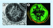
Figure 3.. Association of Coxiella cell envelope projections with the Coxiella-containing vacuole membrane.
(A) Transmission electron micrograph and (B) 3D pseudocolored tomograph of a THP-1 macrophage infected for 48 h with Coxiella. Cell envelope-associated needle-like structures (˜20 nm in length) at points of Coxiella–coxiella-containing vacuole membrane contact (arrows) are evident that are likely components of the T4BSS. Bar =100 nm.

References

    1. Raoult D, Marrie T, Mege J. Natural history and pathophysiology of Q fever. Lancet Infect. Dis. 2005;5:219–226. - PubMed
    1. Kampschreur LM, Delsing CE, Groenwold RH, et al. Chronic Q fever in the Netherlands 5 years after the start of the Q fever epidemic: results from the Dutch chronic Q fever database. J. Clin. Microbiol. 2014;52:1637–1643. - PMC - PubMed
    1. Babudieri C. Q fever: a zoonosis. Adv. Vet. Sci. 1959;5:81–84.
    1. Angelakis E, Raoult D. Q fever. Vet. Microbiol. 2010;140:297–309. - PubMed
    1. Hackert VH, Van Der Hoek W, Dukers-Muijrers N, et al. Q fever: single-point source outbreak with high attack rates and massive numbers of undetected infections across an entire region. Clin. Infect. Dis. 2012;55:1591–1599. - PubMed

Substances

LinkOut - more resources