. 2016 Jul 15;353(6296):295-9.
doi: 10.1126/science.aae0104.

Structural basis for integration of GluD receptors within synaptic organizer complexes

Affiliations

Structural basis for integration of GluD receptors within synaptic organizer complexes

Jonathan Elegheert et al. Science. .

Abstract

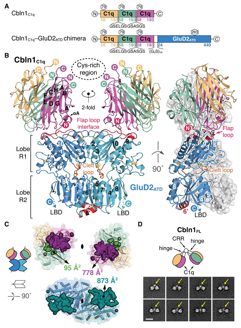
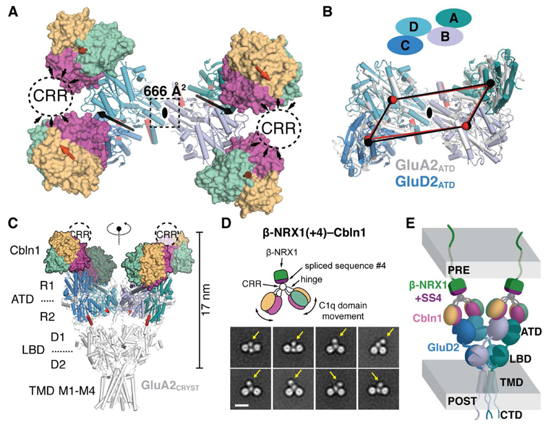
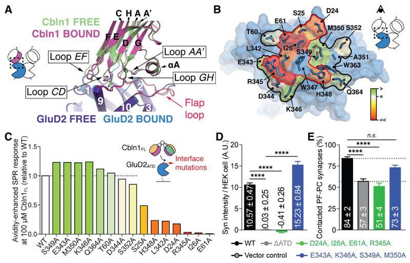
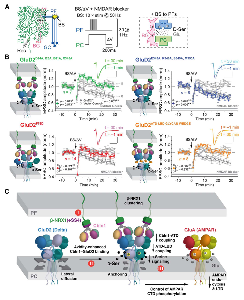
Ionotropic glutamate receptor (iGluR) family members are integrated into supramolecular complexes that modulate their location and function at excitatory synapses. However, a lack of structural information beyond isolated receptors or fragments thereof currently limits the mechanistic understanding of physiological iGluR signaling. Here, we report structural and functional analyses of the prototypical molecular bridge linking postsynaptic iGluR δ2 (GluD2) and presynaptic β-neurexin 1 (β-NRX1) via Cbln1, a C1q-like synaptic organizer. We show how Cbln1 hexamers "anchor" GluD2 amino-terminal domain dimers to monomeric β-NRX1. This arrangement promotes synaptogenesis and is essential for D: -serine-dependent GluD2 signaling in vivo, which underlies long-term depression of cerebellar parallel fiber-Purkinje cell (PF-PC) synapses and motor coordination in developing mice. These results lead to a model where protein and small-molecule ligands synergistically control synaptic iGluR function.


Figures

Fig. 1
Fig. 1. Architecture of the Cbln1C1q–GluD2ATD Binary Complex.
(A) Schematic representation of the chimeric Cbln1C1q and Cbln1C1q–GluD2ATD constructs. (B) “Front” and “side” view of the Cbln1C1q–GluD2ATD complex. The inward tilted orientation of the Cbln1 C1q domains suggests the position of the Cbln1 cysteine-rich region (CRR), as visible in EM class averages and represented by a dotted ellipse here. GluD2 α-helix 6’ and flap and cleft loops are highlighted. Disulfide bridges are shown as yellow spheres. (C) Symmetry-breaking in the Cbln1C1q interface. The total buried interaction surface is shown in a 90° rotated open book view. (D) Selected negative-stain EM class averages of Cbln1FL illustrate its dimer-of-trimers arrangement. Yellow arrows indicate the suggested position of the CRR that links both C1q trimers. Scale bar: 10 nm.
Fig. 2
Fig. 2. Quaternary Structure of the β-NRX1–Cbln1–GluD2 Complex.
(A) “Top” view of the full Cbln1C1q–GluD2ATD dimer-of-dimers complex. The black ellipse, black arrows and red arrows indicate the overall two-fold symmetry axis, the two-fold symmetry axes in the GluD2ATD dimers and the three-fold symmetry axes in the Cbln1C1q trimers, respectively. The suggested position of the Cbln1 CRR is marked with dashed ovals. (B) Superposition of the GluD2 and GluA2 (PDB 3KG2, (5)) N-shaped ATD layers using the B–D ATD dimers (view equivalent to (A)). Centers of mass of GluD2ATD (black spheres) and GluA2ATD (red spheres) are connected to highlight overall similarity. (C) View along the overall two-fold axis of the Cbln1C1q–GluD2ATD complex aligned to Y-shaped GluA2CRYST using the B–D ATD dimers. (D) Selected negative-stain EM class averages of the β-NRX1(+4)–Cbln1FL complex. Yellow arrows indicate the suggested position of β-NRX1(+4). Scale bar; 10 nm. CRR; cysteine-rich region. (E) Model of the synapse-spanning β-NRX1(+4)–Cbln1–GluD2 complex.
Fig. 3
Fig. 3. Details and Structure-guided Mutagenesis of the Cbln1–GluD2 Interface.
(A) Superposition of free and bound Cbln1C1q and GluD2ATD. (B) GluD2 alanine-scanning mutagenesis; SPR response levels are color-annotated as a heat map onto the GluD2 ATD structure. The mutated interface is outlined in black, and the interaction hotspot is outlined in red. (C) GluD2 alanine-scanning mutagenesis using Cbln1FL and monomerised GluD2ATD. The bar chart shows absolute SPR responses after stimulation with 100 µM Cbln1, relative to wild-type GluD2. (D) Quantification of hemi-synapse formation by cerebellar granule cells (GCs) and HEK 293T cells expressing structure-guided GluD2 mutants. Syn; synaptophysin. Data represent mean ± SEM. ****; p < 0.0001 (Kruskal-Wallis and Steel-Dwass test). (E) Quantification of contacted synapses between PFs and PCs expressing structure-guided GluD2 mutants in Grid2-null (GluD2-deficient) cerebella. Data represent mean ± SEM. ****; p < 0.0001, n.s.; not significant (Kruskal-Wallis and Steel-Dwass test).
Fig. 4
Fig. 4. Structure-guided GluD2 Mutants Affect Cerebellar Synaptic Plasticity.
(A) Setup to induce LTD in immature PC dendrites. PC: Purkinje cell, GC: granule cell, PF: parallel fiber, BG: Bergmann glia, BS: burst stimulation, Rec: recording electrode. (B) Averaged LTD data from immature Grid2-null (GluD2-deficient) PCs expressing GFP + GluD2 variants, after burst PF stimulation (BS) combined with direct PC depolarization (ΔV) (BS/ΔV; arrow). The insets show PF-EPSCs at t = –1 min and t = 30 min time points relative to BS/ΔV application. Data represent the mean ± SEM. **; p < 0.01, *; p < 0.05, n.s.; not significant (Kruskal-Wallis and Steel-Dwass test). NMDA receptor blockers are 100 µM D-AP5 plus 25 µM MK801. (C) Proposed key events leading to signal transmission in the β-NRX1(+4)–Cbln1– GluD2 triad. I; β-NRX1–Cbln1 is a pre-synaptic anchor for GluD2. II; trans-synaptic complex formation. GluD2 allows binding of two β-NRX1(+4)–Cbln1 complexes and is shown at full occupancy. III; GluD2, Cbln1 and D-Ser cooperatively induce postsynaptic LTD.

References

    1. Traynelis SF, et al. Glutamate receptor ion channels: structure, regulation, and function. Pharmacological reviews. 2010;62:405–496. doi: 10.1124/pr.109.002451. - DOI - PMC - PubMed
    1. Karakas E, Furukawa H. Crystal structure of a heterotetrameric NMDA receptor ion channel. Science. 2014;344:992–997. doi: 10.1126/science.1251915. - DOI - PMC - PubMed
    1. Lee CH, et al. NMDA receptor structures reveal subunit arrangement and pore architecture. Nature. 2014;511:191–197. doi: 10.1038/nature13548. - DOI - PMC - PubMed
    1. Nakagawa T, Cheng Y, Ramm E, Sheng M, Walz T. Structure and different conformational states of native AMPA receptor complexes. Nature. 2005;433:545–549. doi: 10.1038/nature03328. - DOI - PubMed
    1. Sobolevsky AI, Rosconi MP, Gouaux E. X-ray structure, symmetry and mechanism of an AMPA-subtype glutamate receptor. Nature. 2009;462:745–756. doi: 10.1038/nature08624. - DOI - PMC - PubMed

Publication types