. 2016 Aug 2;113(31):E4494-503.
doi: 10.1073/pnas.1608423113. Epub 2016 Jul 14.

Severe muscle wasting and denervation in mice lacking the RNA-binding protein ZFP106

Affiliations

Severe muscle wasting and denervation in mice lacking the RNA-binding protein ZFP106

Douglas M Anderson et al. Proc Natl Acad Sci U S A. .

Abstract

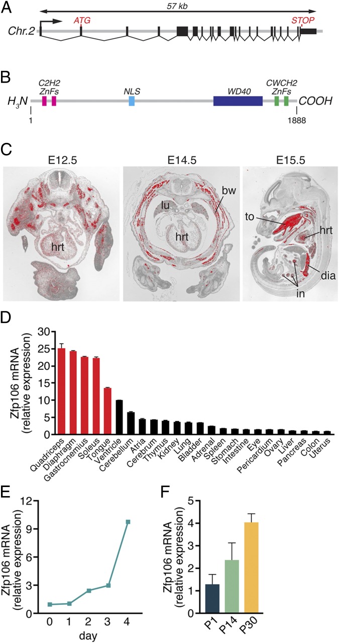
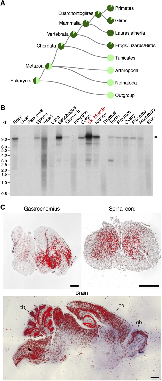
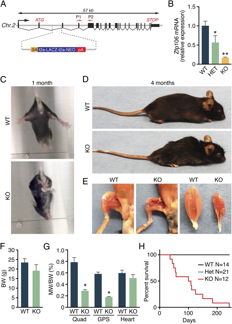
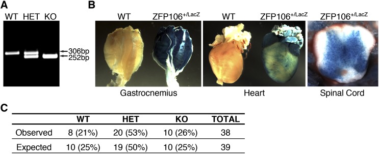
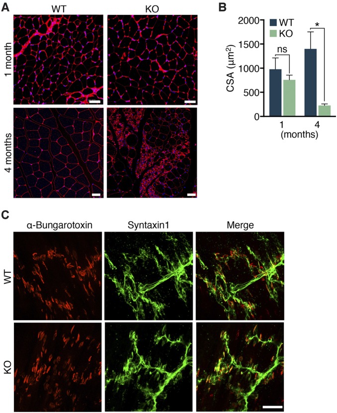
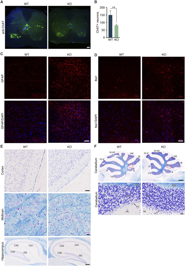
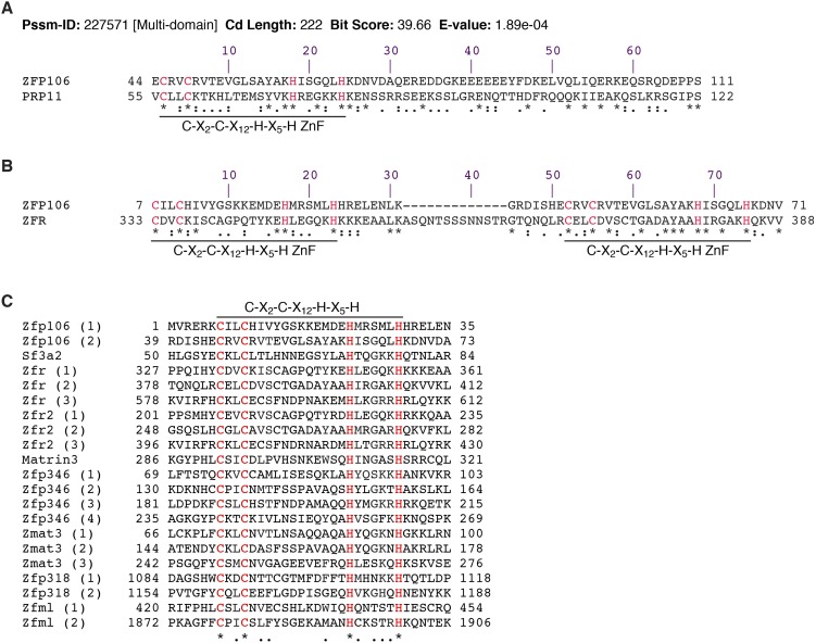
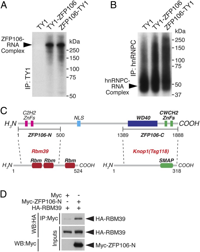
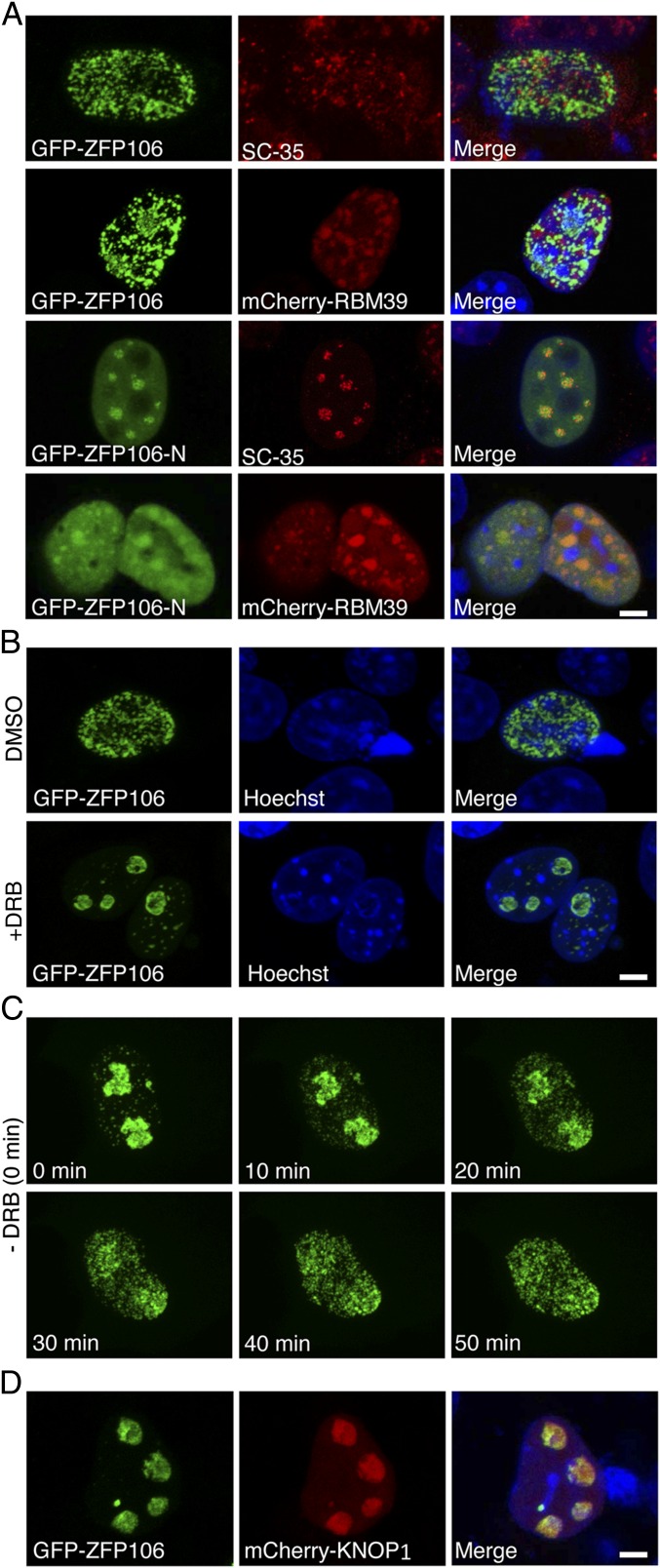
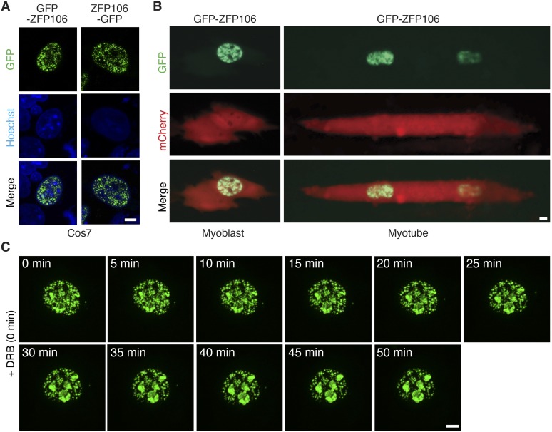
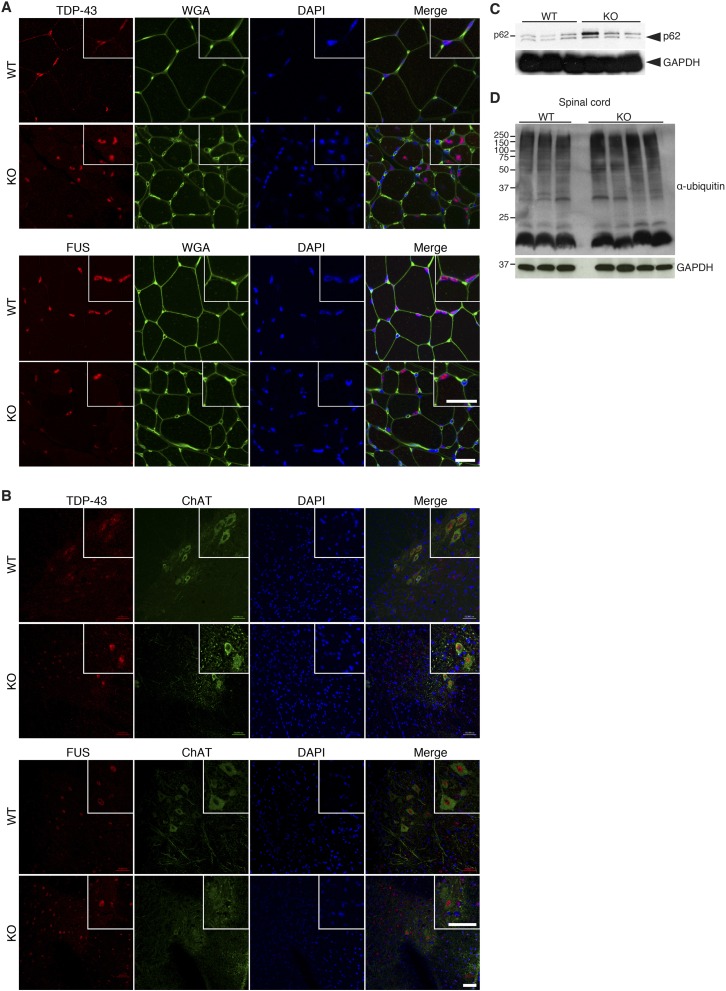
Innervation of skeletal muscle by motor neurons occurs through the neuromuscular junction, a cholinergic synapse essential for normal muscle growth and function. Defects in nerve-muscle signaling cause a variety of neuromuscular disorders with features of ataxia, paralysis, skeletal muscle wasting, and degeneration. Here we show that the nuclear zinc finger protein ZFP106 is highly enriched in skeletal muscle and is required for postnatal maintenance of myofiber innervation by motor neurons. Genetic disruption of Zfp106 in mice results in progressive ataxia and hindlimb paralysis associated with motor neuron degeneration, severe muscle wasting, and premature death by 6 mo of age. We show that ZFP106 is an RNA-binding protein that associates with the core splicing factor RNA binding motif protein 39 (RBM39) and localizes to nuclear speckles adjacent to spliceosomes. Upon inhibition of pre-mRNA synthesis, ZFP106 translocates with other splicing factors to the nucleolus. Muscle and spinal cord of Zfp106 knockout mice displayed a gene expression signature of neuromuscular degeneration. Strikingly, altered splicing of the Nogo (Rtn4) gene locus in skeletal muscle of Zfp106 knockout mice resulted in ectopic expression of NOGO-A, the neurite outgrowth factor that inhibits nerve regeneration and destabilizes neuromuscular junctions. These findings reveal a central role for Zfp106 in the maintenance of nerve-muscle signaling, and highlight the involvement of aberrant RNA processing in neuromuscular disease pathogenesis.

Keywords: ZNF106; amyotrophic lateral sclerosis (ALS); motor neuron disease (MND); neuromuscular junction (NMJ); pre-mRNA splicing.


Conflict of interest statement

The authors declare no conflict of interest.

Figures

Fig. 1.
Fig. 1.
Zfp106 gene structure and expression. (A) The mouse Zfp106 gene is located on chromosome 2 and is composed of 21 exons, spanning ∼57 kb. (B) The mouse ZFP106 protein is 1,888 amino acids long and contains a bipartite nuclear localization signal (NLS), four zinc fingers, and a WD40 domain. (C) Section in situ hybridization using a probe antisense to Zfp106 showed strong expression in skeletal muscle, heart, intestine, and lungs during embryogenesis. bw, body wall muscle; dia, diaphragm; hrt, heart; in, intestine; lu, lung; to, tongue. (D) The expression of Zfp106 in adult mouse tissues was measured using qRT-PCR. Zfp106 transcript expression was highest in fast- and slow-type skeletal muscles (red bars), heart, and brain. Detectable levels of Zfp106 transcripts were broadly expressed in the adult tissues examined. (E and F) Zfp106 expression, quantified using qRT-PCR, increased during C2C12 muscle cell differentiation in vitro (E) and in gastrocnemius–plantaris muscles isolated from P1, P14, and P30 mice (F). Mean ± SD.
Fig. S1.
Fig. S1.
Conservation and expression of Zfp106. (A) Phylogram generated by Treefam (22) showing that Zfp106 orthologs are found in diverse vertebrate species, including mammals, birds, reptiles, amphibians, and fish. (B) Northern blot analysis of adult mouse tissues using a probe specific to Zfp106 showed enrichment in skeletal muscle, esophagus, kidney, colon, and brain. The arrow marks the predicted transcript size of the full-length Zfp106 transcript encoding the 1,888-aa ZFP106 protein. (C) Section in situ hybridization on adult mouse tissues using an antisense probe to Zfp106 showing expression in gastrocnemius, spinal cord, and brain. cb, cerebellum; ce, cerebral cortex; ob, olfactory bulb. (Scale bars, 1 mm.)
Fig. 2.
Fig. 2.
Postnatal muscle wasting in Zfp106 KO mice. (A) Diagram showing the Zfp106 LacZ knock-in allele obtained from the KOMP Repository. P1 and P2 indicate the locations of Zfp106 qRT-PCR primers. (B) Zfp106 transcript levels were reduced by 85% in mice homozygous for the LacZ allele (KO), measured by qRT-PCR in gastrocnemius–plantaris muscles. (C) At ∼1 mo of age, Zfp106 KO mice display abnormal hindlimb clasping. (D) At 4 mo of age, Zfp106 KO mice are approximately the same size as their WT littermates but show kyphotic curvature of the spine. (E) Gross examination of hindlimb skeletal muscles of 4-mo-old Zfp106 KO mice showed severe muscle wasting. (F and G) At 4 mo of age, no differences were detected in body weight or heart weight of Zfp106 KO and WT mice. Skeletal muscle weight was significantly reduced in Zfp106 KO mice. BW, body weight; GPS, gastrocnemius–plantaris–soleus muscle group; MW, muscle weight; Quad, quadriceps muscle. (H) Kaplan–Meier survival plot showing premature lethality of Zfp106 KO mice. *P < 0.01; **P < 0.0001; Mean ± SD.
Fig. S2.
Fig. S2.
Characterization of Zfp106 KO mice. (A) Duplex PCR-based genotyping strategy used to identify wild-type (WT), heterozygous (HET), and knockout (KO) mice for the Zfp106 LacZ knock-in allele. (B) Staining of adult tissues heterozygous for the Zfp106 LacZ knock-in allele showed β-galactosidase staining in skeletal muscles, heart, and spinal cord. (C) Number of mice born versus expected from heterozygous intercrosses of Zfp106 LacZ knock-in mice.
Fig. 3.
Fig. 3.
Severe wasting and denervation in Zfp106 KO muscle. (A) Transverse sections of gastrocnemius muscles from 1- and 4-mo-old WT and Zfp106 KO mice were stained with Masson’s trichrome. Electron micrographs (EM) of WT and KO gastrocnemius revealed disorganized sarcomere structures in 4-mo-old muscles from Zfp106 KO muscle. (Scale bars, 1 μm.) (B) Gastrocnemius muscle from 1- and 4-mo-old WT and Zfp106 KO mice were stained with anti-Syntaxin 1 (green) and Texas red-conjugated α-bungarotoxin (red) to assess neuromuscular junctions. White arrowheads indicate end plates that lack nerve terminal juxtaposition in Zfp106 KO gastrocnemius muscles. (Scale bars, for 1-mo WT and KO mice, 20 μm; for 4-mo WT and KO mice, 50 μm.)
Fig. S3.
Fig. S3.
Progressive skeletal muscle atrophy in Zfp106 KO mice. (A and B) Wheat germ agglutinin (red) and DAPI (blue) staining of transverse myofibers of 1- and 4-mo-old Zfp106 KO mice showed an 80% decrease in myofiber cross-sectional area (CSA) in Zfp106 KO gastrocnemius muscle. *P < 0.01. ns, not significant. Mean ± SD. (Scale bars, 20 μm.) (C) Motor axons and nerve terminals were stained using an antibody against Syntaxin 1 (green) and Texas red-conjugated α-bungarotoxin to label the motor end plates at the neuromuscular junctions (NMJs). At postnatal day 1 (P1), no differences in NMJ structure, number, or innervation by motor nerves were detected in gastrocnemius muscles of Zfp106 KO compared with WT littermates. (Scale bar, 50 μm.)
Fig. 4.
Fig. 4.
Motor axon atrophy and degeneration in Zfp106 KO mice. (A) Light microscopy of toluidine blue-stained transverse sections of sciatic nerves isolated from WT and Zfp106 KO mice at 2 wk and 4 mo of age. (Scale bar, 100 µm.) (B) Axon morphology in sciatic nerves shown in A. The yellow arrowhead points to a degenerating axon in a Zfp106 KO sciatic nerve section. (Scale bar, 20 µm.) (C) Electron microscopy analysis showed degenerating motor axons present in the sciatic nerve of Zfp106 KO mice. Yellow arrowheads point to degenerating motor axons. SC, Schwann cell nucleus. (Scale bar, 2 µm.)
Fig. S4.
Fig. S4.
Peripheral neurodegeneration in Zfp106 KO mice. (A and B) Transverse sections through the lumbar spinal cord of WT and Zfp106 KO mice stained with an antibody against choline acetyltransferase (ChAT) showed a 43% decrease in the number of ChAT+ motor neurons at 2 mo of age. **P < 0.01. Mean ± SD. (Scale bar, 100 μm.) (C and D) Immunohistochemistry using antibodies specific to GFAP and Iba1 in the lumbar spinal cord revealed an increase in the number of active astrocytes and microglia cells in Zfp106 KO mice compared with WT littermates, respectively. (Scale bars, 50 μm.) (E and F) Nissl/Luxol fast blue staining of brain tissues revealed no overt differences in the morphology, myelination, or number of neurons in 3-mo-old Zfp106 KO mice compared with WT littermates. Red box shows the magnified area below. [Scale bars, 50 μm (cortex), 20 μm (midbrain), 200 μm (hippocampus), 500 μm (cerebellum; Top), and 20 μm (cerebellum; Bottom).] DG, dentate gyrus; GL, granular layer; ML, molecular layer; PC, Purkinje cells; WM, white matter.
Fig. S5.
Fig. S5.
Sequence homology between ZFP106 and RNA-binding ZnF proteins. (A) A search of the Conserved Domain Database using the amino acid sequence of ZFP106 identified homology to the zinc finger splicing factor PRP11, a homolog of mammalian SF3A2. Protein alignment shows that the region encompasses an N-terminal ZnF of ZFP106 and ZnF of PRP11. (B) The N-terminal ZnFs of ZFP106 share significant homology with the ZnFs of zinc finger RNA-binding protein (ZFR). (C) Alignment of the ZnFs of ZFP106 and other RNA-binding ZnF proteins that contain the conserved C2H2 ZnF motif C-X2-C-X12-H-X5-H. Cysteine and histidine residues comprising the C-X2-C-X12-H-X5-H motif are highlighted in red. “*” denotes identical, “:” denotes strongly similar and “.” denotes weakly similar residues.
Fig. 5.
Fig. 5.
ZFP106 is an RNA-binding protein that interacts with RBM39. (A) Autoradiograph resolving the ZFP106–RNA complex after cross-linking and immunoprecipitation (IP) of TY1 epitope-tagged ZFP106 in Neuro2a cells. (B) Immunoprecipitation using a primary antibody against hnRNPC was used as a control for RNA integrity. (C) Diagram showing the ZFP106 fragments used for a yeast two-hybrid screen of a mouse normalized cDNA library. The 500-aa N-terminal fragment (ZFP106-N) and the 500-aa C-terminal fragment (ZFP106-C) identified RBM39 and KNOP1 as ZFP106-interacting proteins, respectively. SMAP, small acidic protein. (D) Western blot (WB) analysis showing that HA epitope-tagged RBM39 coprecipitated with a Myc epitope-tagged fragment of the N-terminal ZFP106 fragment (Myc–ZFP106-N) in COS7 cells.
Fig. 6.
Fig. 6.
Nuclear localization and translocation of ZFP106. (A) Confocal microscopy of COS7 cells showing coexpression and localization of GFP-ZFP106 with SC-35 or mCherry-RBM39. The N-terminal fragment of ZFP106 (ZFP106-N) localized to a nuclear speckle pattern that overlapped with SC-35 and an mCherry-RBM39 fusion protein. (B) Inhibition of total cellular RNA synthesis by DRB treatment induced the translocation of GFP-ZFP106 from nuclear speckles to the nucleolus. (C) After removal of DRB, GFP-ZFP106 regained a nuclear speckle pattern over the course of 50 min. (D) Coexpression of GFP-ZFP106 and mCherry-KNOP1 induced the translocation of ZFP106 from nuclear speckles to the nucleolus. (Scale bars, 5 µm.)
Fig. S6.
Fig. S6.
Subcellular localization of ZFP106 in COS7 and C2C12 muscle cells. (A) Confocal microscopy of transiently transfected COS7 cells showed GFP-ZFP106 and ZFP106-GFP localized to nuclear speckles. (B) The nuclear speckle pattern of GFP-ZFP106 was also found in both C2C12 myoblasts and myotubes. Coexpression of an unlocalized mCherry fluorescent protein was used to denote cell shape. (Scale bars, 5 µm.) (C) Inhibition of RNA synthesis by DRB treatment of COS7 cells transfected with GFP-ZFP106. A panel of fluorescence micrographs acquired by live-cell imaging every 5 min over the course of 50 min showed GFP-ZFP106 progressively localized to nucleoli.
Fig. 7.
Fig. 7.
Gene profiling and alternative splicing changes in Zfp106 KO tissues. (A) Genes up-regulated and down-regulated in both skeletal muscle and spinal cord of 1-mo-old Zfp106 KO mice. (B) Quantitative real-time PCR for a subset of genes dysregulated in Zfp106 KO tissues. *P < 0.05. ND, not detected. (C) Quantitative real-time PCR showing expression of Nogo and Nogo-A transcript levels in WT and Zfp106 KO gastrocnemius–plantaris muscles at 1 mo of age. *P < 0.01. (D) Robust up-regulation of NOGO-A protein was observed in gastrocnemius–plantaris muscle of 1-mo-old Zfp106 KO mice, detected using an antibody specific to the A isoform of Nogo. Western blot analysis for GAPDH was used as a loading control. Mean ± SD.
Fig. S7.
Fig. S7.
Protein processing and degradation in Zfp106 KO mice. (A and B) Immunohistochemistry using antibodies specific to TDP-43 and FUS revealed normal subcellular localization in skeletal muscle (A) and spinal cord (B) of 3-mo-old Zfp106 KO mice compared with WT littermates. WGA, wheat germ agglutinin. Insets are magnification showing stained cell nuclei. (Scale bars, A, 50 μm; B, 25 μm.) (C) Western blot analysis revealed similar levels of p62 protein in spinal cord from Zfp106 KO and WT littermate mice. (D) Normal levels of polyubiquitinated proteins were detected in Zfp106 KO spinal cord lysates using an antibody against ubiquitin.

References

    1. Hirsch NP. Neuromuscular junction in health and disease. Br J Anaesth. 2007;99(1):132–138. - PubMed
    1. Goulet BB, Kothary R, Parks RJ. At the “junction” of spinal muscular atrophy pathogenesis: The role of neuromuscular junction dysfunction in SMA disease progression. Curr Mol Med. 2013;13(7):1160–1174. - PubMed
    1. Moloney EB, de Winter F, Verhaagen J. ALS as a distal axonopathy: Molecular mechanisms affecting neuromuscular junction stability in the presymptomatic stages of the disease. Front Neurosci. 2014;8:252. - PMC - PubMed
    1. Pollari E, Goldsteins G, Bart G, Koistinaho J, Giniatullin R. The role of oxidative stress in degeneration of the neuromuscular junction in amyotrophic lateral sclerosis. Front Cell Neurosci. 2014;8:131. - PMC - PubMed
    1. Tintignac LA, Brenner HR, Rüegg MA. Mechanisms regulating neuromuscular junction development and function and causes of muscle wasting. Physiol Rev. 2015;95(3):809–852. - PubMed

Publication types

MeSH terms

Substances