. 2016 Jun 27:10:1167-74.
doi: 10.2147/OPTH.S103443. eCollection 2016.

Effects of anti-vascular endothelial growth factor monoclonal antibody (bevacizumab) on lens epithelial cells

Affiliations

Effects of anti-vascular endothelial growth factor monoclonal antibody (bevacizumab) on lens epithelial cells

Jong Hwa Jun et al. Clin Ophthalmol. .

Abstract

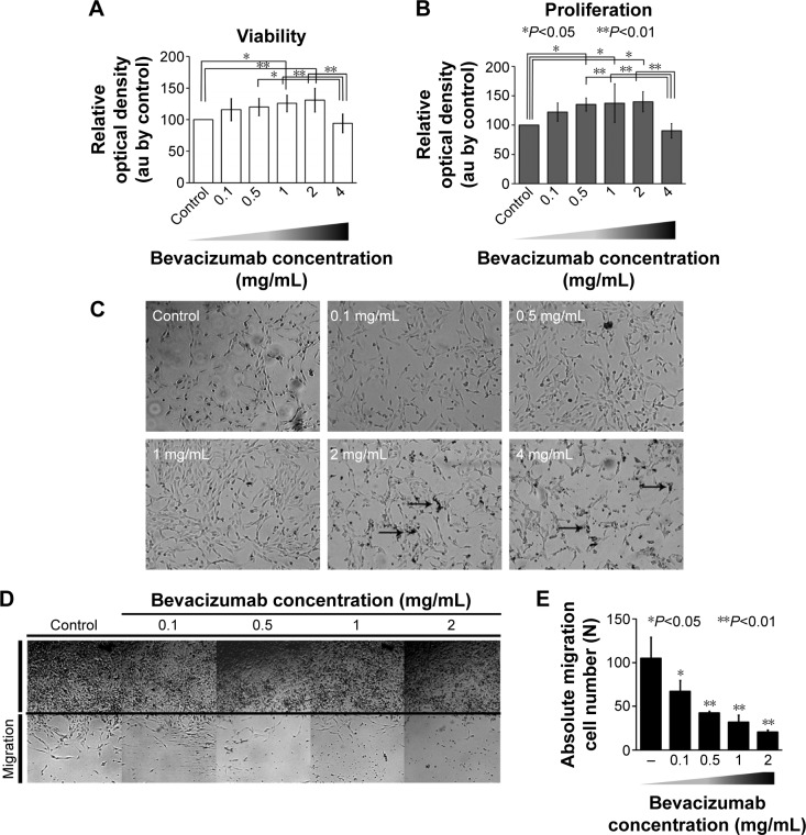
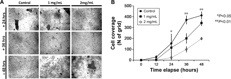
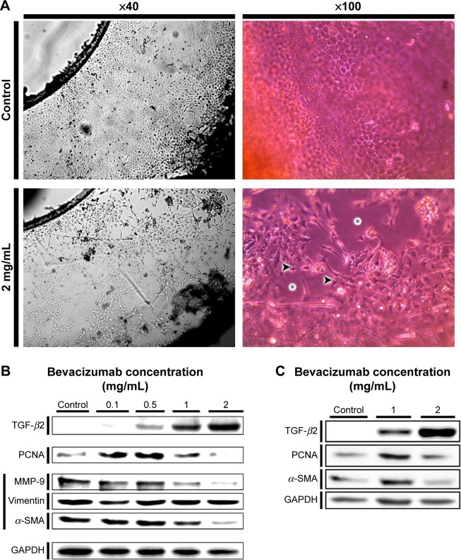
The molecular and cellular effects of anti-vascular endothelial growth factor monoclonal antibody (bevacizumab) on lens epithelial cells (LECs) were examined using both an immortalized human lens epithelial cell line and a porcine capsular bag model. After treatment with various concentrations of bevacizumab, cell viability and proliferation patterns were evaluated using the water-soluble tetrazolium salt assay and 5-bromo-2'-deoxyuridine enzyme-linked immunosorbent assay, respectively. The scratch assay and Western blot analysis were employed to validate the cell migration pattern and altered expression levels of signaling molecules related to the epithelial-mesenchymal transition (EMT). Application of bevacizumab induced a range of altered cellular events in a concentration-dependent manner. A 0.1-2 mg/mL concentration demonstrated dose-dependent increase in proliferation and viability of LECs. However, 4 mg/mL decreased cell proliferation and viability. Cell migrations displayed dose-dependent retardation from 0.1 mg/mL bevacizumab treatment. Transforming growth factor-β2 expression was markedly increased in a dose-dependent manner, and α-smooth muscle actin, matrix metalloproteinase-9, and vimentin expression levels showed dose-dependent changes in a B3 cell line. Microscopic observation of porcine capsular bag revealed changes in cellular morphology and a decline in cell density compared to the control after 2 mg/mL treatment. The central aspect of posterior capsule showed delayed confluence, and the factors related to EMT revealed similar expression patterns to those identified in the cell line. Based on these results, bevacizumab modulates the proliferation and viability of LECs and induces morphological alterations through the modulation of expression patterns of specific factors related to the EMT.

Keywords: avastin; bevacizumab; lens epithelial cell; transforming growth factor; vascular endothelial growth factor.


Figures

Figure 1
Figure 1
Effects of bevacizumab on proliferation and migration of LECs. Notes: (A) Cell viability assay using the cell counting kit-8 after treatment with various bevacizumab concentrations. In LECs, treatment with 4 mg/mL bevacizumab showed a significant decrease in cell viability compared with control, 0.1 mg/mL, 0.5 mg/mL, 1 mg/mL, and 2 mg/mL bevacizumab treatment after 72-hour incubation. Up to 2 mg/mL bevacizumab, cell viability increased in a dose-dependent manner. (B) BrdU enzyme-linked immunosorbent proliferation assay after treatment with various bevacizumab concentrations. As in the cell viability assay, cell proliferation increased with bevacizumab dose up to 2 mg/mL concentration but declined abruptly at 4 mg/mL concentration. (C) Microscopic images after application of various bevacizumab concentrations. In human lens epithelial B3 cells, treatment with 4 mg/mL bevacizumab showed a significant decline in cell density compared with control, 0.1 mg/mL, 0.5 mg/mL, 1 mg/mL, and 2 mg/mL bevacizumab treatment after 72-hour incubation. The densities of viable attached cells increased in a dose-dependent manner, but 4 mg/mL bevacizumab treatment resulted in an abrupt decrement in cell density. Interestingly, treatment with 1 mg/mL or 2 mg/mL bevacizumab showed a significant number of detached nonviable cells in the culture medium, but the attached cell density was higher than for cells treated with lower concentrations (0 mg/mL, 0.1 mg/mL, and 0.5 mg/mL). These results agreed with the BrdU proliferation and cell viability assays. Arrowheads indicate shrunken cells (magnification, 40×). (D and E) Scratch assay for cell migration after treatment with various bevacizumab concentrations and 72-hour treatment under cell starvation conditions. Microscopic images show that LEC migrations from the scratch edges decreased in a dose-dependent manner. Migration of LECs statistically significantly decreased at the lowest dose of bevacizumab (0.1 mg/mL; P<0.05, Tukey’s HSD post hoc test, by oneway-ANOVA). The inhibitory effect on migration was apparent over 0.5 mg/mL bevacizumab compared with control (P<0.01, Tukey’s HSD post hoc test, by oneway-ANOVA). Magnification, ×40. Results are in response to a one way analysis of variance (ANOVA). *P<0.05, **P<0.01. Abbreviations: LECs, lens epithelial cells; BrdU, 5-bromo-2′-deoxyuridine; HSD, honest significant difference; ANOVA, analysis of variance.
Figure 2
Figure 2
Central confluence of posterior capsule in porcine lenses. (A) Central coverage of posterior capsule decreased in a dose-dependent manner (B) After treatment with various doses of bevacizumab, coverage of the posterior capsule in porcine lenses. Notes: After treatment with various doses of bevacizumab, coverage of the posterior capsule decreased in the first 12 hours at 2 mg/mL bevacizumab (P<0.05, by one way-ANOVA). In addition, with an increase of incubation time, the control group showed a greater difference in cell coverage than both 1 mg/mL and 2 mg/mL groups at 36 hours (P<0.01, Tukey’s HSD post hoc test, by oneway-ANOVA) and 48 hours (P<0.05 and P<0.01 in 1 mg/mL and 2 mg/mL, respectively, Tukey’s HSD post hoc test, by oneway-ANOVA). Data are displayed as mean ± SD. Magnification, ×40. Results are in response to a one way analysis of variance (ANOVA). *P<0.05, **P<0.01. Abbreviations: HSD, honest significant difference; h, hours; ANOVA, analysis of variance.
Figure 3
Figure 3
Effects of bevacizumab on cellular morphology and expression of factors related to EMT and proliferation. Notes: (A) Microscopic morphology of anterior capsular LECs for control and 2 mg/mL bevacizumab treatment in lens capsular bag after 120 hours cultivation. Capsular bag LECs showed decreased cell density (asterisks) and bleb-like morphological changes after cultivation in 2 mg/mL bevacizumab (arrowheads). (B and C) Western blot analysis after treatment with various bevacizumab concentrations in cultured cell and porcine lens capsular bags. TGF-β2 is markedly increased in a dose-dependent manner in both systems. Western blot analysis of cultured cells revealed increase-peak-decrease patterns in PCNA and α-SMA expression around 0.5 mg/mL concentration. The expression pattern of PCNA is in agreement with results from the cell viability assay by cell counting kit-8 and the BrdU proliferation assay. Overall, MMP-9 showed a decrease in expression over 1 mg/mL treatment, whereas vimentin expression increased in a dose-dependent manner. Similarly, PCNA and α-SMA expression levels peaked at 1 mg/mL bevacizumab in the porcine lens capsular bag model. Abbreviations: LECs, lens epithelial cells; TGF-β2, transforming growth factor-β2; PCNA, proliferating cell nuclear antigen; α-SMA, α-smooth muscle actin; BrdU, 5-bromo-2′-deoxyuridine; MMP-9, matrix metalloproteinase-9; GAPDH, glyceraldehyde-3-phosphate dehydrogenase.

Similar articles

Cited by

References

    1. McNulty R, Wang H, Mathias RT, Ortwerth BJ, Truscott RJ, Bassnett S. Regulation of tissue oxygen levels in the mammalian lens. J Physiol. 2004;559(pt 3):883–898. - PMC - PubMed
    1. Neelam S, Brooks MM, Cammarata PR. Lenticular cytoprotection. Part 1: the role of hypoxia inducible factors-1alpha and -2alpha and vascular endothelial growth factor in lens epithelial cell survival in hypoxia. Mol Vis. 2013;19:1–15. - PMC - PubMed
    1. Shui YB, Wang X, Hu JS, et al. Vascular endothelial growth factor expression and signaling in the lens. Invest Ophthalmol Vis Sci. 2003;44(9):3911–3919. - PubMed
    1. Saint-Geniez M, Kurihara T, D’Amore PA. Role of cell and matrix-bound VEGF isoforms in lens development. Invest Ophthalmol Vis Sci. 2009;50(1):311–321. - PMC - PubMed
    1. Pennock S, Kim D, Mukai S, et al. Ranibizumab is a potential prophylaxis for proliferative vitreoretinopathy, a nonangiogenic blinding disease. Am J Pathol. 2013;182(5):1659–1670. - PMC - PubMed

LinkOut - more resources