

Calreticulin mediates an invasive breast cancer phenotype through the transcriptional dysregulation of p53 and MAPK pathways
- PMID: 27418879
- PMCID: PMC4944499
- DOI: 10.1186/s12935-016-0329-y
Calreticulin mediates an invasive breast cancer phenotype through the transcriptional dysregulation of p53 and MAPK pathways
Abstract
Background: The introduction of effective novel biomarkers of invasion and metastasis is integral for the advancement of breast cancer management. The present study focused on the identification and evaluation of calreticulin (CRT) as a potential biomarker for breast cancer invasion.
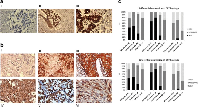
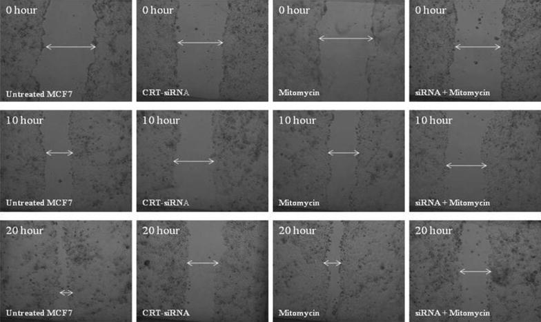
Methods: Two-dimensional gel protein electrophoresis and MALDI-TOF were utilized in the analysis of fresh-frozen invasive intra-ductal carcinoma specimens. Calreticulin-associated expression was analyzed using immunohistochemistry of FFPE non-malignant/malignant breast specimens. A CRT-knockdown model of MCF7 cell line was developed using siRNA and the CRT genotype/phenotype correlations based on migration and trans-well invasion assays were determined. Finally, microarray-based global gene expression profiling was conducted to elucidate the possible calreticulin pro-invasive regulatory pathways.
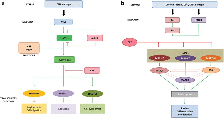
Results: Two-dimensional gel protein electrophoresis and MALDI-TOF analysis showed upregulation of calreticulin expression in tumor tissues as compared to the normal adjacent tissues. Meta-analysis of the immunohistochemical results confirmed significantly higher expression of calreticulin (p < 0.05) in the stromal compartments of malignant tissues as compared to non-malignant tissues. Migration and transwell invasion assays showed significant loss in the migratory and invasive potential of CRT-knockdown cells (p < 0.05). Global gene expression profiling successfully identified various putative gene networks such as p53 and MAPK pathways that are involved in calreticulin breast cancer signaling.
Conclusion: Besides confirming calreticulin overexpression in invasive breast cancer tissues, this study reveals a calreticulin-dependent pro-invasive potential and suggests possible contributing pathways. Defining the mechanistic role of invasion and characterizing the possible calreticulin-dependent molecular targets will be the focus of future work.
Keywords: Breast cancer; Calreticulin; Invasion; MAPK; p53.
Figures


Similar articles
-
Identification of calreticulin as a prognosis marker and angiogenic regulator in human gastric cancer.Ann Surg Oncol. 2009 Feb;16(2):524-33. doi: 10.1245/s10434-008-0243-1. Epub 2008 Dec 3. Ann Surg Oncol. 2009. PMID: 19050968
-
Calreticulin promotes migration and invasion of esophageal cancer cells by upregulating neuropilin-1 expression via STAT5A.Clin Cancer Res. 2014 Dec 1;20(23):6153-62. doi: 10.1158/1078-0432.CCR-14-0583. Epub 2014 Sep 17. Clin Cancer Res. 2014. PMID: 25231404
-
Overexpression of calreticulin contributes to the development and progression of pancreatic cancer.J Cell Physiol. 2014 Jul;229(7):887-97. doi: 10.1002/jcp.24519. J Cell Physiol. 2014. PMID: 24264800
-
Calreticulin as a marker and therapeutic target for cancer.Clin Exp Med. 2023 Sep;23(5):1393-1404. doi: 10.1007/s10238-022-00937-7. Epub 2022 Nov 6. Clin Exp Med. 2023. PMID: 36335525 Review.
-
Calreticulin and cancer.Pathol Oncol Res. 2013 Apr;19(2):149-54. doi: 10.1007/s12253-012-9600-2. Epub 2013 Feb 8. Pathol Oncol Res. 2013. PMID: 23392843 Review.
Cited by
-
Role of Nerve Growth Factor (NGF) and miRNAs in Epithelial Ovarian Cancer.Int J Mol Sci. 2017 Feb 26;18(3):507. doi: 10.3390/ijms18030507. Int J Mol Sci. 2017. PMID: 28245631 Free PMC article. Review.
-
Dexamethasone-Mediated Upregulation of Calreticulin Inhibits Primary Human Glioblastoma Dispersal Ex Vivo.Int J Mol Sci. 2018 Feb 14;19(2):572. doi: 10.3390/ijms19020572. Int J Mol Sci. 2018. PMID: 29443896 Free PMC article.
-
High expression of calreticulin indicates poor prognosis and modulates cell migration and invasion via activating Stat3 in nasopharyngeal carcinoma.J Cancer. 2019 Aug 29;10(22):5460-5468. doi: 10.7150/jca.35362. eCollection 2019. J Cancer. 2019. PMID: 31632490 Free PMC article.
-
Immunostimulatory Properties of Chemotherapy in Breast Cancer: From Immunogenic Modulation Mechanisms to Clinical Practice.Front Immunol. 2022 Jan 5;12:819405. doi: 10.3389/fimmu.2021.819405. eCollection 2021. Front Immunol. 2022. PMID: 35069604 Free PMC article. Review.
-
Proteome derangement in malignant epithelial cells and its stroma following exposure to 4-(methylnitrosamino)-1-(3-pyridyl)-1-butanone.Arch Toxicol. 2023 Mar;97(3):711-720. doi: 10.1007/s00204-022-03426-8. Epub 2022 Nov 25. Arch Toxicol. 2023. PMID: 36434399 Free PMC article.
References
-
- Berns EMJJ. Use of micro array analysis to predict out come and response in breast cancer patients. Br J Cancer Suppl. 2004;9:49–50.
-
- Green M, Raina V. Epidemiology, screening and diagnosis of breast cancer in the Asia-Pacific region: current perspectives and important considerations. Asia-Pacific J Clin Oncol. 2008;4:S5–S13. doi: 10.1111/j.1743-7563.2008.00191.x. - DOI
LinkOut - more resources
Full Text Sources
Other Literature Sources
Molecular Biology Databases
Research Materials
Miscellaneous
