. 2016 Mar;11(1):97-102.
doi: 10.1177/1558944715614854. Epub 2016 Jan 14.

Elbow Reconstruction With Compression Plate Arthrodesis and Circumferential Muscle-Sparing Latissimus Dorsi Flap After Tumor Resection: A Case Report

Affiliations

Elbow Reconstruction With Compression Plate Arthrodesis and Circumferential Muscle-Sparing Latissimus Dorsi Flap After Tumor Resection: A Case Report

Zhi Yang Ng et al. Hand (N Y). 2016 Mar.

Abstract

Background: The goals of limb-sparing surgery in the setting of extremity malignancies are 2-fold: oncological clearance and the rehabilitation of function and aesthetics. Treatment success should be defined by the extent of restoration of the patient's premorbid function for reintegration into society.

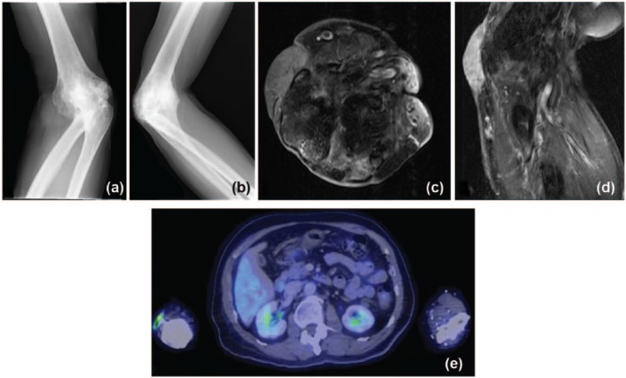
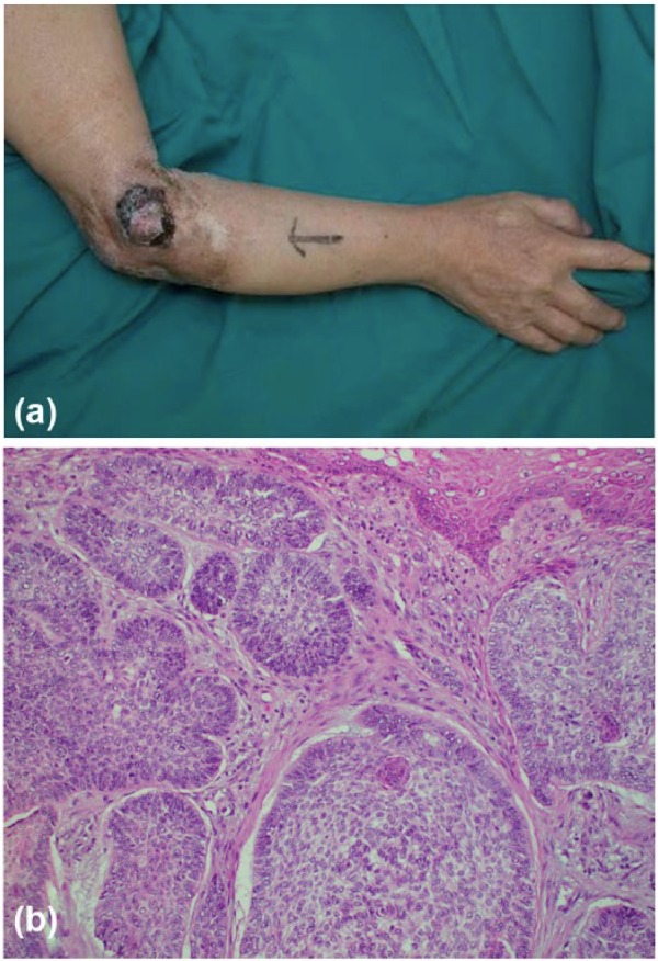
Methods: We would like to report an unusual case of a patient with a chronically ankylosed elbow with joint invasion by basal cell carcinoma which resulted from malignant transformation of an overlying, long-standing wound due to inadequately treated septic arthritis from his childhood years.

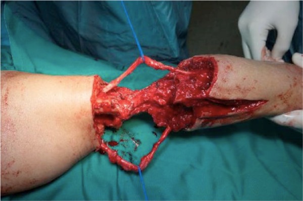
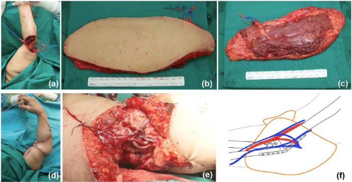
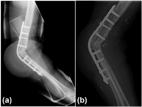
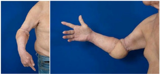
Results: Following R0 resection, upper limb shortening and compression plate elbow arthrodesis were performed with the aim of restoring the degree of upper limb function that the patient had been accustomed to preoperatively. The resultant circumferential defect was then closed with a contralateral, free muscle-sparing latissimus dorsi flap.

Conclusions: Functional preservation may therefore be more important than the mere restoration of anatomical defects in these especially challenging situations.

Keywords: arthrodesis; elbow; free flap; limb salvage; upper extremity.


Conflict of interest statement

Declaration of Conflicting Interests: The author(s) declared no potential conflicts of interest with respect to the research, authorship, and/or publication of this article.

Figures

Figure 1.
Figure 1.
Preoperative imaging studies: (a, b) X-rays showed marked cortical irregularities with sclerosis of articulating bones, disorganization and arthrodesis of the proximal radioulnar and radiocapitellar joints; (c, d) T1-weighted MRI with contrast revealed almost complete ankylosis of the elbow with hypointensity suggestive of calcification at the radiocapitellar joint, subcutaneous soft-tissue edema over the olecranon, and diffuse skin thickening with a heterogeneous slightly hyperintense and enhancing lesion at the lateral aspect of the elbow; (e) PET-CT was only significant for focal FDG uptake in the lateral aspect of the right elbow. Note. MRI, magnetic resonance imaging; PET-CT, positron emission tomography–computed tomography; FDG, fluorodeoxyglucose.
Figure 2.
Figure 2.
(a) Preoperative clinical view of lesion over posterolateral aspect of right ankylosed elbow. (b) Histopathology demonstrates peripheral palisading of the tumor cells with stromal retraction clefting characteristic of basal cell carcinoma (hematoxylin-eosin, original magnification ×200).
Figure 3.
Figure 3.
Postexcision circumferential defect including exposure of the radial and ulnar nerves, and transected basilic and cephalic veins (not shown).
Figure 4.
Figure 4.
(a) Postexcision circumferential defect with exposure of the radial and ulnar nerves and plate after limb shortening and joint arthrodesis; (b, c) harvest of a 24×10-cm muscle-sparing free latissimus dorsi flap; (d) flap inset in a 360° wrap-around fashion; and microsurgical anastomosis (e) intraoperative and (f) schematic views. Note. TDA was anastomosed end to side to the BA; the TDV and a branch of the SAV were both anastomosed end to end to the CV and a branch of the BV, respectively, via interpositional SSV grafts. TDA, thoracodorsal artery; BA, brachial artery; TDV, thoracodorsal vein; SAV, serratus anterior vein; CV, cephalic vein; BV, basilic vein; SSV, short saphenous vein.
Figure 5.
Figure 5.
Postoperative x-rays: (a) at 9 months showing early signs of callous formation; (b) at 23 months follow-up with evidence of bony union across the site of joint arthrodesis.
Figure 6.
Figure 6.
Functional preservation achieved with satisfactory restoration of the patient’s preoperative upper limb to its preoperative state. Note. Clinical deformity seen is due to morbidity arising from oncological resection leading to partial loss of flexor and extensor muscle origins; satisfactory regain of hand function was achieved with hand therapy.

Similar articles

Cited by

References

    1. Anwar U, Al Ghazal SK, Ahmad M, Sharpe DT. Horrifying basal cell carcinoma forearm lesion leading to shoulder disarticulation. Plast Reconstr Surg. 2006;117:6e-9e. - PubMed
    1. Bray PW, Bell RS, Bowen CV, Davis A, O’Sullivan B. Limb salvage surgery and adjuvant radiotherapy for soft tissue sarcomas of the forearm and hand. J Hand Surg Am. 1997;22:495-503. - PubMed
    1. Chun JM, Byeon HK. Shoulder arthrodesis with a reconstruction plate. Int Orthop. 2009;33:1025-1030. - PMC - PubMed
    1. Koller H, Kolb K, Assuncao A, Kolb W, Holz U. The fate of elbow arthrodesis: indications, techniques, and outcome in fourteen patients. J Shoulder Elbow Surg. 2008;17:293-306. - PubMed
    1. Kopp J, Polykandriotis E, Loos B, et al. Giant rodent ulcer of the elbow requiring defect coverage by preconditioned latissimus dorsi pedicled myocutaneous flap following excision. J Eur Acad Dermatol Venereol. 2007;21:252-254. - PubMed

LinkOut - more resources