

Ten-Year Experience of Cutaneous and/or Subcutaneous Infections Due to Coelomycetes in France
- PMID: 27419178
- PMCID: PMC4943527
- DOI: 10.1093/ofid/ofw106
Ten-Year Experience of Cutaneous and/or Subcutaneous Infections Due to Coelomycetes in France
Abstract
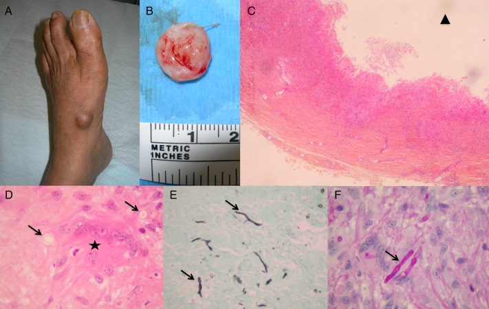
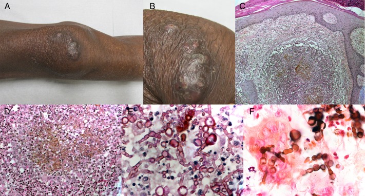
Background. Coelomycetes are rarely but increasingly reported in association with human infections involving mostly skin and subcutaneous tissues, both in immunocompetent and immunocompromised patients. Coelomycetes constitute a heterogeneous group of filamentous fungi with distinct morphological characteristics in culture, namely an ability to produce asexual spores within fruit bodies. Methods. We included all cases of proven primary cutaneous and/or subcutaneous infections due to coelomycetes received for identification at the French National Reference Center for Invasive Mycoses and Antifungals between 2005 and 2014. Eumycetoma, chromoblastomycosis, and disseminated infections were excluded. Results. Eighteen cases were analyzed. The median age was 60.5 years. In all cases, patients originated from tropical or subtropical areas. An underlying immunodepression was present in 89% of cases. Cutaneous and/or subcutaneous lesions, mainly nodules, abscesses, or infiltrated plaques, were observed in distal body areas. Isolates of different genera of coelomycetes were identified: Medicopsis (6), Paraconiothyrium (3), Gloniopsis (3), Diaporthe (3), Peyronellaea (2), Lasiodiplodia (1). Lesion treatment consisted of complete (10) or partial (2) surgical excision and/or the use of systemic antifungal therapy, namely voriconazole (5) and posaconazole (4). Literature review yielded 48 additional cases of cutaneous and/or subcutaneous infections due to coelomycetes. Conclusions. Infectious diseases physicians should suspect coelomycetes when observing cutaneous and/or subcutaneous infections in immunocompromised hosts from tropical areas; a sequence-based approach is crucial for strains identification but must be supported by consistent phenotypic features; surgical treatment should be favored for solitary, well limited lesions; new triazoles may be used in case of extensive lesions, especially in immunocompromised patients.
Keywords: Medicopsis romeroi; Paraconiothyrium sp; coelomycetes; cutaneous phaeohyphomycosis; subcutaneous abscess.
Figures

References
-
- Stchigel A, Sutton D. Coelomycete fungi in the clinical lab. Current Fungal Infection Reports 2013; 7:171–91.
-
- Garcia-Reyne A, López-Medrano F, Morales JM et al. Cutaneous infection by Phomopsis longicolla in a renal transplant recipient from Guinea: first report of human infection by this fungus. Transpl Infect Dis 2011; 13:204–7. - PubMed
-
- Ajello L, Georg LK, Steigbigel RT, Wang CJ. A case of phaeohyphomycosis caused by a new species of Phialophora. Mycologia 1974; 66:490–8. - PubMed
-
- McGinnis MR. Chromoblastomycosis and phaeohyphomycosis: new concepts, diagnosis, and mycology. J Am Acad Dermatol 1983; 8:1–16. - PubMed
LinkOut - more resources
Full Text Sources
Other Literature Sources
