. 2016 Nov 1;23(11):1827-1838.
doi: 10.1038/cdd.2016.69. Epub 2016 Jul 15.

Cell death is not essential for caspase-1-mediated interleukin-1β activation and secretion

Affiliations

Cell death is not essential for caspase-1-mediated interleukin-1β activation and secretion

S A Conos et al. Cell Death Differ. .

Abstract

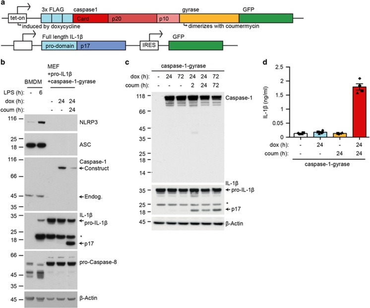
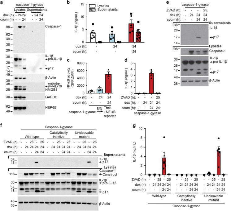
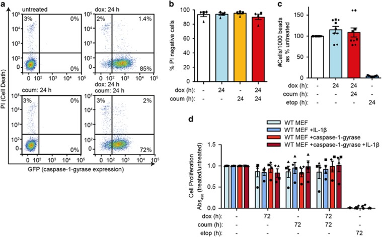
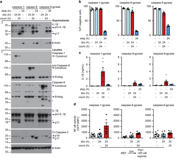
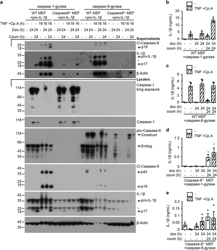
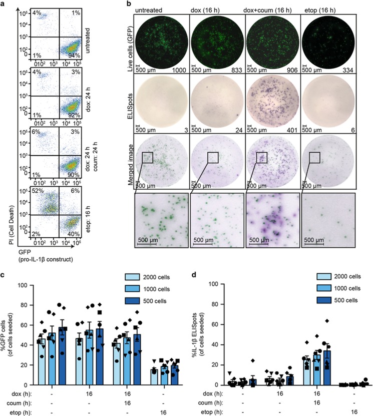
Caspase-1 cleaves and activates the pro-inflammatory cytokine interleukin-1 beta (IL-1β), yet the mechanism of IL-1β release and its dependence on cell death remains controversial. To address this issue, we generated a novel inflammasome independent system in which we directly activate caspase-1 by dimerization. In this system, caspase-1 dimerization induced the cleavage and secretion of IL-1β, which did not require processing of caspase-1 into its p20 and p10 subunits. Moreover, direct caspase-1 dimerization allowed caspase-1 activation of IL-1β to be separated from cell death. Specifically, we demonstrate at the single cell level that IL-1β can be released from live, metabolically active, cells following caspase-1 activation. In addition, we show that dimerized or endogenous caspase-8 can also directly cleave IL-1β into its biologically active form, in the absence of canonical inflammasome components. Therefore, cell death is not obligatory for the robust secretion of bioactive IL-1β.


Figures

Figure 1
Figure 1
Forced dimerization of caspase-1 causes IL-1β cleavage in the absence of an intact inflammasome pathway. (a) A schematic of the doxycycline inducible fusion protein FLAG-caspase-1-gyrase-GFP (caspase-1-gyrase) vector system and pro-IL-1β retroviral vector system. Doxycycline treatment induces expression of caspase-1-gyrase fusion protein, whereas coumermycin binds to the gyrase domain to cause caspase-1 dimerization. (b) MEFs lack expression of the inflammasome components NLRP3, ASC and caspase-1. WT BMDMs were treated with 100 ng/ml LPS for 6 h to induce NLRP3 expression. WT MEFs, previously infected with caspase-1-gyrase and a pro-IL-1β, were treated with 1 μg/ml doxycycline to induce caspase-1-gyrase expression and 700 nM coumermycin to dimerize caspase-1-gyrase for the indicated times. Cells lysates were analyzed by western blot for the indicated proteins. (c) Dimerization of caspase-1-gyrase with coumermycin caused rapid cleavage of pro-IL-1β. Western blot of lysates from MEFs containing caspase-1-gyrase and pro-IL-1β, which were treated with 1 μg/ml doxycycline and 700 nM coumermycin for the indicated times. Blot is representative of three independent experiments. (d) Dimerization of caspase-1-gyrase causes secretion of IL-1β. MEFs stably infected with caspase-1-gyrase vector and pro-IL-1β were treated with 1 μg/ml doxycycline and 700 nM coumermycin. Supernatants were analyzed by ELISA. n=4 independent experiments. Error bars represent the S.E.M. Asterisks denote non-specific bands in western blots
Figure 2
Figure 2
Biologically active IL-1β is secreted from MEFs following caspase-1 dimerization. (a and b) Dimerization of caspase-1-gyrase causes cleavage and secretion of IL-1β. MEFs stably infected with caspase-1-gyrase vector and pro-IL-1β were treated with 1 μg/ml doxycycline and 700 nM coumermycin. Lysates and supernatants were adjusted to be of equal volume when harvested. (a) Equal volumes of lysates and supernatants were analyzed by western blot. Blot is representative of three independent experiments. (b) One-third of cleaved cellular IL-1β is released into the supernatant. IL-1β detected from equal volumes of lysates and supernatants by ELISA. n=5 independent experiments. (c) IL-1β secreted from MEFs is biologically active. Supernatants (S/N) from MEFs stably infected with caspase-1-gyrase and pro-IL-1β, treated for 24 h with 1 μg/ml doxycycline and 700 nM coumermycin, were transferred onto Thp1 cells bearing an NF-κB GFP reporter construct. After 24 h incubation, GFP expression was quantified by flow cytometry. Data are expressed as the change in mean fluorescence intensity (MFI) relative to Thp1 cells in media alone. n=3 independent experiments. (d and e) Secretion of IL-1β following caspase-1 activation requires catalytic caspase activity. MEFs containing pro-IL-β and caspase-1-gyrase were treated with 1 μg/ml doxycycline, 700 nM coumermycin and 25 μM pan-caspase inhibitor Z-VAD-fmk for the indicated times. Supernatants were analyzed by (d) ELISA (n=3 independent experiments) and (e) western blot, whereas lysates were analyzed by (e) western blot. (f and g) Cleavage and secretion of IL-1β requires catalytic activity of caspase-1 but not auto-processing. MEFs stably infected with pro-IL-1β and wild-type caspase-1-gyrase, or a catalytically inactive mutant (C385G), or an uncleavable mutant (D296N, D300N, D304N, D308N, D313N, D314N) were treated with 1 μg/ml doxycycline, 700 nM coumermycin and 25 μM Z-VAD-fmk. (f) Lysates were analyzed by western blot, whereas supernatants were analyzed by (f) western blot and (g) ELISA. n=3-5 independent experiments. Error bars represent the S.E.M. in all graphs. Asterisks denote non-specific bands in western blots. See also Supplementary Figure S1 and S2
Figure 3
Figure 3
Dimerization of caspase-1-gyrase in MEFs does not induce cell death. (a and b) Caspase-1-gyrase was induced in MEFs with 1 μg/ml doxycycline and dimerized with 700 nM coumermycin for the indicated times. Cell viability was measured by PI uptake and flow cytometric analysis. (a) Representative flow cytometry data. (b) Quantification of cell viability from four independent experiments. (c) Dimerization of caspase-1-gyrase does not alter the total number of viable cells. MEFs containing caspase-1-gyrase were treated with 1 μg/ml doxycycline and 700 nM coumermycin, or 34 μM etoposide as specified for the indicated times and analyzed by flow cytometry. The absolute number of PI-negative cells was measured as a ratio to unstained flow cytometry beads. n=3 independent experiments, each performed in triplicate. (d) Cell proliferation and metabolism is not altered by caspase-1-gyrase dimerization. MEFs containing caspase-1-gyrase and pro-IL-1β constructs were treated as in (c) for the indicated times and cellular proliferation was measured by the MTT-PMS viability assay. n=4 independent experiments. Error bars represent the S.E.M. in all graphs
Figure 4
Figure 4
Caspase-8-gyrase dimerization can induce low levels of IL-1β secretion. (a) Caspase-8-gyrase dimerization causes IL-1β cleavage and secretion. Western blot analysis of lysates and supernatants from MEFs containing overexpressed pro-IL-1β and either caspase-1-gyrase, caspase-8-gyrase or caspase-9-gyrase treated as specified with 1 μg/ml doxycycline and 700 nM coumermycin, or 34 μM etoposide, for the indicated times. Asterisks denote non-specific bands. (b) Caspase-8-gyrase and caspase-9-gyrase dimerization induces small amounts of cell death. Cells were treated as in (a) and analyzed by flow cytometry for uptake of PI. n=4 independent experiments. (c) IL-1β is secreted upon dimerization and activation of caspases. MEFs were treated as in (a) and IL-1β released into the supernatant was measured by ELISA. n=4–6 independent experiments. (d) IL-1β secreted following caspase-1-gyrase or caspase-8-gyrase dimerization is biologically active. Thp1 cells bearing an NF-κB GFP reporter were incubated for 24 h with supernatants from caspase-1-gyrase, caspase-8-gyrase or caspase-9-gyrase MEFs treated for 24 h with 1 μg/ml doxycycline and 700 nM coumermycin. GFP expression indicative of NF-κB activation was quantified as the change in mean fluorescence intensity (MFI) relative to Thp1 cells in media alone. n=6 independent experiments. Error bars represent the S.E.M. in all graphs. Asterisks denote non-specific bands in western blots. Data for caspase-1, -8 and -9 were collected in parallel. See also Supplementary Figure S3
Figure 5
Figure 5
Both endogenous caspase-8 and caspase-8-gyrase can cleave IL-1β and cause its secretion. MEFs containing pro-IL-1β and either caspase-1-gyrase (a, b and d) or caspase-8-gyrase (a, c and e) vectors were treated with 1 μg/ml doxycycline and 700 nM coumermycin, as well as 100 ng/ml TNF and 1 μM Cp.A when indicated by cross-hatched columns. (a) Representative western blot of lysates and supernatants blotted for the indicated proteins. Asterisks denote non-specific bands. (b, c, d and e) Activation of endogenous caspase-8, caspase-8-gyrase or caspase-1-gyrase causes secretion of IL-1β. Supernatant of indicated cells was analyzed by IL-1β ELISA after treatment. Error bars represent S.E.M. n=3–5 independent experiments. See also Supplementary Figure S4
Figure 6
Figure 6
IL-1β is secreted from live MEFs when caspase-1-gyrase is dimerized. (a) GFP fluorescence is lost upon cell death. MEF cells containing pro-IL-1β and caspase-1-gyrase were treated with 1 μg/ml doxycycline and 700 nM coumermycin, or 34 μM etoposide for indicated times, and GFP fluorescence and PI uptake were analyzed by flow cytometry. (b, c and d) Caspase-1 activity causes secretion of IL-1β from live cells. MEF cells containing pro-IL-1β and caspase-1-gyrase constructs were seeded at three densities on the ELISpot plate. After 16 h treatment with 1 μg/ml doxycycline and 700 nM coumermycin, or 34 μM etoposide, GFP images were taken and then ELISPot staining was immediately performed. (b) Representative images of 2000 cells/well ELISpot experiment. The brightness and contrast of images were adjusted using Fiji (ImageJ) and Adobe Illustrator in a linear fashion, with settings applied equally to every image. GFP images were inverted in the merged image and pseudo-coloring was applied using Adobe Illustrator to allow overlay of ELISpots and GFP-positive cells. (c) Analysis of the number of GFP-positive cells as a ratio of total cells seeded. (d) Analysis of ELISpots represented as a percentage of cells seeded per well. Both GFP and ELISpot images were acquired and quantified using an AID ELISpot Reader. n=6 independent experiments. Error bars represent the S.E.M. See also Supplementary Figure S5

References

    1. Menu P, Vince JE. The NLRP3 inflammasome in health and disease: the good, the bad and the ugly. Clin Exp Immunol 2011; 166: 1–15. - PMC - PubMed
    1. Vince JE. When beauty is skin deep: regulation of the wound response by caspase-8, RIPK3, and the inflammasome. J Invest Dermatol 2015; 135: 1936–1939. - PubMed
    1. Lawlor KE, Vince JE. Ambiguities in NLRP3 inflammasome regulation: is there a role for mitochondria? Biochim Biophys Acta 2014; 1840: 1433–1440. - PubMed
    1. Monteleone M, Stow JL, Schroder K. Mechanisms of unconventional secretion of IL-1 family cytokines. Cytokine 2015; 74: 213–218. - PubMed
    1. Vince JE, Silke J. The intersection of cell death and inflammasome activation. Cell Mol Life Sci 2016; 73: 2349–2367. - PMC - PubMed

LinkOut - more resources