. 2016 Aug 16;7(33):53459-53470.
doi: 10.18632/oncotarget.10557.

Gelsolin suppresses gastric cancer metastasis through inhibition of PKR-p38 signaling

Affiliations

Gelsolin suppresses gastric cancer metastasis through inhibition of PKR-p38 signaling

Xiangliang Yuan et al. Oncotarget. .

Abstract

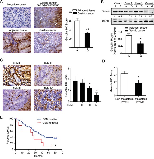
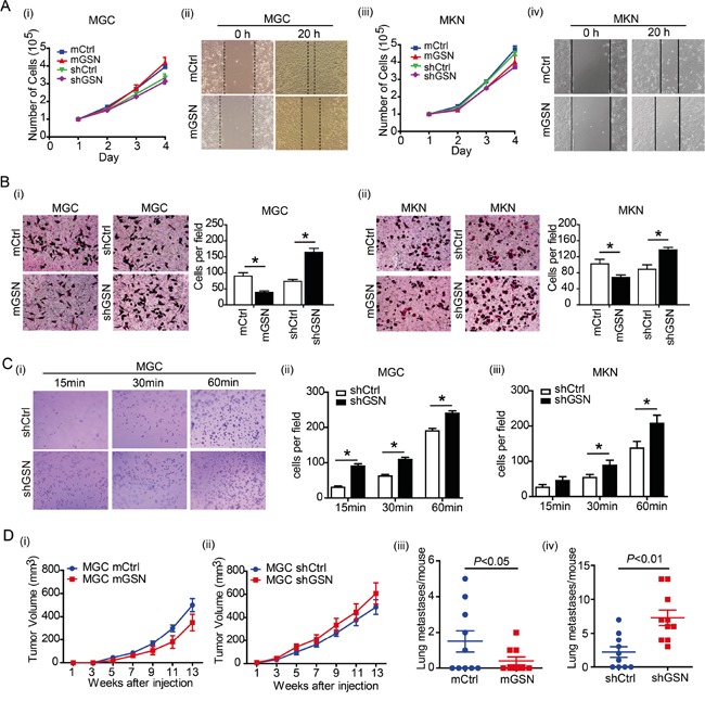
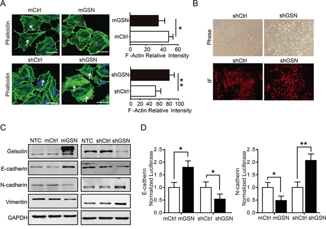
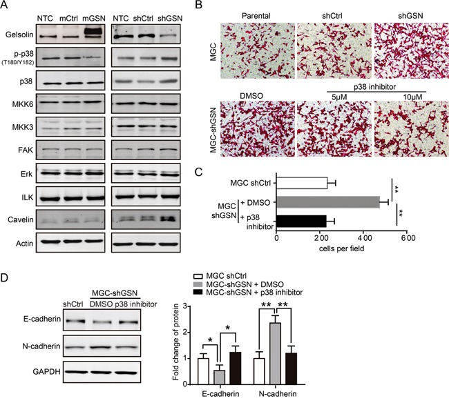
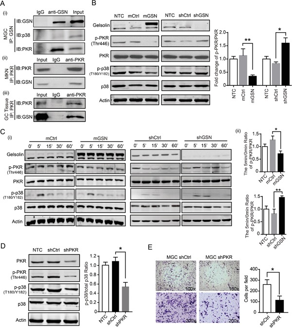
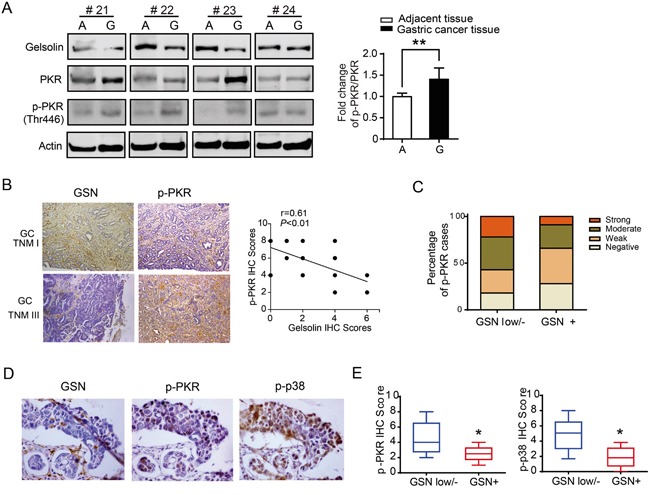
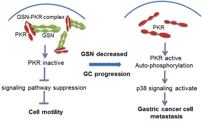
The biological function of gelsolin in gastric cancer and its mechanism remained undefined. Here, we demonstrated that gelsolin was down-regulated in human gastric cancer tissues, and lower tumorous gelsolin significantly correlated with gastric cancer metastasis. Functionally, gelsolin suppressed the migration of gastric cancer cells in vitro and inhibited lung metastasis in vivo. In mechanism, gelsolin decreased epithelial-mesenchymal transition (EMT) inducing cytoskeleton remolding through inhibition of p38 signaling to suppress the migration of gastric cancer cell. Moreover, gelsolin bound to and decreased the phosphorylation of PKR, and then inhibited p38 signaling pathway. Finally, similar to the gastric cancer cell lines, PKR-p38 signaling pathway proteins tend to be activated and correlated with low expression of gelsolin in clinical gastric cancer tissues. Altogether, these results highlight the importance of gelsolin in suppression of gastric cancer metastasis through inhibition of PKR-p38 signaling pathway.

Keywords: PKR; gastric cancer; gelsolin; metastasis; p38MAPK protein kinase.


Conflict of interest statement

The authors have no conflict of interest to declare.

Figures

Figure 1
Figure 1. Downregulated gelsolin negatively correlated with tumor stage in gastric cancer
A. Immunohistochemistry (IHC) staining of gelsolin in human paraffin-embedded gastric cancer and adjacent tissues. IgG1 was used for the negative control of gelsolin (upper left, 400x). Representative IHC image of gelsolin in gastric cancer (arrow) and adjacent tissues (arrowhead) (upper right, 200x). Magnified image showed the different expression pattern of gelsolin in adjacent tissues (bottom left, 400x) and gastric cancer (bottom right, 400x). Statistical analyses of the average score of gelsolin staining between GC tissues (G) and corresponding adjacent tissues in 20 cases (A). (**, P<0.01). B., immunoblotting (IB) analysis of gelsolin expression in 4 cases of human gastric cancer tissues (G) and matched adjacent tissues (A). Quantification of IB analyses for gelsolin in 20 cases of matched human tissues was conducted (*, P<0.05).C., Representative IHC images of gelsolin expression in different stages of gastric cancer tissues (TNM I, II, III, IV) (400x).(*, P<0.05 compared to TNM-I). D., Statistical analyses of average score of gelsolin according to the metastasis status (*, P<0.05). E., Kaplan–Meier survival curve analyses of 56 patients with gastric cancer determined by IHC scoring of gelsolin as negative (score=0) versus positive (score >0). (*, P<0.05).
Figure 2
Figure 2. Gelsolin suppress the migration of gastric cancer cells in vitro and in vivo
A., The cell proliferation of MGC (i) or MKN (iii) transfected with over-expression (mGSN) or knockdown (shGSN) of gelsolin. Wound healing assay carried out with MGC (ii) or MKN (iv) with gelsolin overexpression. Representative images of triplicate experiments were shown. B., Transwell assay carried out with MGC (i) or MKN (ii) with over-expression (mGSN) or knockdown (shGSN) of gelsolin. Three independent experiments with 3 fields for each were performed, and the representative fields were shown (*, P<0.05, student's t-test). C., Cell adhesion assay was performed in MGC (i, ii) or MKN cell (iii) transfected with shCtrl or shGSN after 15, 30, and 60 minutes. Bar graph represents mean ±s.d. of quantification of adherent cell numbers at indicated time point (n=3; *,P<0.05, student's t-test). D., Tumor size measurement of mice transplanted with GSN-overexpressed MGC cells (mGSN or control) (i), or GSN-knockdown MGC cells (shGSN or control)(ii) (n=10 per group). Results are expressed as mean ± SEM. After 13 weeks, mice were euthanized and the numbers of metastases in the lungs were counted (iii and iv).
Figure 3
Figure 3. Gelsolin inhibited EMT and actin cytoskeletal rearrangement to suppress the migration of gastric cancer cell
A., Immunofluorescence staining for phalloidin (F-actin) in MGC cell with overexpression (mGSN) or knockdown (shGSN) of gelsolin. Scar bar, 10μm. White arrows showed the filamentous F-actin. Quantitative results of the actin fluorescence intensity were presented as mean ± SEM of triplicate experiments (*, P<0.05; **, P<0.01). B., Morphology of MGC cell stably transfected with shCtrl or shGSN was examined by phase contrast microscopy and immunofluorescence (RFP). C., Immunoblotting analyses of EMT-related proteins in MGC cell with overexpression or knockdown gelsolin. D., Luciferase activity assay of E-cadherin or N-cadherin promoter-driven luciferase expression in indicated cells after 48h post-transfection (*, P<0.05; **, P<0.01).
Figure 4
Figure 4. Gelsolin repressed EMT by inhibiting p38 pathway
A., Immunoblotting analysis of migration-related proteins in MGC cell with over-expression or knockdown of gelsolin. B., Transwell assay carried out with indicated MGC cells with or without the treatment of described concentration of p38 inhibitor. C., Statistical analyses of migratory cells in indicated MGC cells (**,P<0.01). D., Immunoblotting analyses of EMT markers (E-cadherin, N-cadherin) in indicated MGC cells with control (DMSO) or p38 inhibitor (10 μM). Bar graph represents mean±s.d. of western blot quantification of E-cadherin and N-cadherin expression (n=3; *,P<0.05; **,P<0.01, student's t-test).
Figure 5
Figure 5. Gelsolin interacted with PKR to inhibitp38 signaling pathway
A., (i) Immunoprecipitation (IP) of gelsolin and immunoblotting (IB) analysis of gelsolin, PKR and p38 in MGC cell. MGC cell lysates were run as an input control, and isotype control IgG1 of gelsolin were run in the left lane. Total cell lysates were prepared from MKN cells (ii) or gastric cancer tissue from one patient with TNM stage-I gastric cancer (iii). Following immunoprecipitation with the anti-PKR antibody or IgG control antibody, total lysates and bound proteins were subjected to immunoblotting with anti-gelsolin antibody. B., Western blot analyses of gelsolin, p-PKR, PKR, p-p38, and p38 proteins in indicated MGC cells. Quantification of fold change of p-PKR/PKR ratio was shown (n=3; *, P<0.05, student's t-test). C., (i) Western blot analyses of p-PKR/total PKR and p-p38/total p38 from indicated MGC cells. After overnight serum starvation, the indicated MGC cells were treated with10% FBS for indicated time points and then lysed for analysis by Western blot. Representative Western blot images are shown.(ii) Bar graph represents the fold change of p-PKR/PKR at 5min time point quantified and normalized against 0 min in indicated MGC cells. (n=3; *, P<0.05; **, P<0.01, student's t-test). D., Western blot analyses of p-p38/total p38 in MGC cells with shCtrl or shPKR for 48h. The fold change of p-p38/p38 in MGC cells with shCtrl or shPKR were quantified and normalized against untreated control cell. Values are means ± standard error means of three independent samples (*, P<0.05, student's t-test). E., Transwell assay carried out with MGC cell transfected knockdown of PKR. Representative Transwell images and statistical data were shown (n=3; *, P<0.05, student's t-test).
Figure 6
Figure 6. Downregulated gelsolin was associated with increased expression of PKR-p38 signaling proteins in human gastric cancer tissues
A., Representative Western blot images of gelsolin, PKR and pPKR protein levels in 4 cases of human gastric cancer tissue (G) and matched adjacent tissue (A) were shown. Bar graph represents mean ±s.d. of western blot quantification of p-PKR/PKR in gastric cancer tissues and matched adjacent tissues (n=20; *, P<0.05, student's t-test). B., Representative IHC image of gelsolin and p-PKR from TNM I case and TNM III case were shown (100x), and the correlation between gelsolin and p-PKR in 20 cases of gastric cancer tissues was analyzed. C., The percentage of p-PKR expression was shown in gastric cancer patients with lower/negative or positive gelsolin expression(P<0.05; x2 test). D., Representative IHC images of gelsolin, p-PKR, and p-p38 from consecutive gastric cancer tissue sections were shown (400x). E., the box and whisker plots showed IHC scores of p-PKR and p-p38 with various gelsolin expression intensities (*, P<0.05).
Figure 7
Figure 7. Proposed model for the mechanism of gelsolin signaling pathway in gastric cancer cell metastasis
In non-metastatic gastric cancer cells, gelsolin interacts with PKR and inhibits the activation of PKR. However, in metastatic cancer cells, gelsolin is down-regulated, which allows for the autophosphorylation of PKR. PKR activation leads to the activation of p38 protein and downstream signaling pathway to promote the metastasis of gastric cancer.

Similar articles

Cited by

References

    1. Jemal A, Bray F, Center MM, Ferlay J, Ward E, Forman D. Global cancer statistics. CA Cancer J Clin. 2011;61:69–90. - PubMed
    1. Siegel R, Ma J, Zou Z, Jemal A. Cancer statistics, 2014. CA Cancer J Clin. 2014;64:9. - PubMed
    1. Zeng H, Zheng R, Guo Y, Zhang S, Zou X, Wang N, Zhang L, Tang J, Chen J, Wei K, Huang S, Wang J, Yu L, et al. Cancer survival in China, 2003-2005: a population-based study. Int J Cancer. 2015;136:1921–1930. - PubMed
    1. Thiery JP. Epithelial-mesenchymal transitions in tumour progression. Nat Rev Cancer. 2002;2:442–454. - PubMed
    1. Nurnberg A, Kitzing T, Grosse R. Nucleating actin for invasion. Nat Rev Cancer. 2011;11:177–187. - PubMed