. 2016 Jul 14;166(2):451-467.
doi: 10.1016/j.cell.2016.06.011.

Mapping the Pairwise Choices Leading from Pluripotency to Human Bone, Heart, and Other Mesoderm Cell Types

Affiliations

Mapping the Pairwise Choices Leading from Pluripotency to Human Bone, Heart, and Other Mesoderm Cell Types

Kyle M Loh et al. Cell. .

Abstract

Stem-cell differentiation to desired lineages requires navigating alternating developmental paths that often lead to unwanted cell types. Hence, comprehensive developmental roadmaps are crucial to channel stem-cell differentiation toward desired fates. To this end, here, we map bifurcating lineage choices leading from pluripotency to 12 human mesodermal lineages, including bone, muscle, and heart. We defined the extrinsic signals controlling each binary lineage decision, enabling us to logically block differentiation toward unwanted fates and rapidly steer pluripotent stem cells toward 80%-99% pure human mesodermal lineages at most branchpoints. This strategy enabled the generation of human bone and heart progenitors that could engraft in respective in vivo models. Mapping stepwise chromatin and single-cell gene expression changes in mesoderm development uncovered somite segmentation, a previously unobservable human embryonic event transiently marked by HOPX expression. Collectively, this roadmap enables navigation of mesodermal development to produce transplantable human tissue progenitors and uncover developmental processes. VIDEO ABSTRACT.


Figures

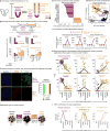
Figure 1
Figure 1. Formation of human primitive streak and its bifurcation into paraxial and lateral mesoderm
A. Each lineage step labeled with a circled number, corresponding to respective sections in the main text and Fig. 7a B. FACS of MIXL1-GFP hESC (Davis et al., 2008) after 24 hrs in anterior or mid PS induction (left); all cells coexpress BRACHYURY and MIXL1 by scRNA-seq; each dot depicts a single cell (right) C. BMP induces, whereas WNT inhibits, lateral mesoderm from the PS on D1-2. (i) qPCR of D1 PS treated with BMP4 or a BMP inhibitor (DM3189) for 24 hours (in the context of A8301 + FGF2); (ii) qPCR of D1 PS treated with WNT agonists (CHIR99021 or WNT3A) or WNT inhibitors (300 ng/mL Dkk1 or 1 μM C59) for 24 hrs (in the context of A8301 + BMP4 [AB]); error bars = S.E.M. for this and all other qPCR experiments D. BMP inhibits, whereas WNT induces, paraxial mesoderm from the PS on D1-2. (i) qPCR of D1 PS treated with BMP4 or a BMP inhibitor (DM3189) for 24 hrs (in the context of A8301 + FGF2 [AF]); (ii) qPCR of D1 PS treated with WNT agonist (3 μM CHIR99021) or WNT inhibitors (2 μM IWR1, 1 μM XAV939 or C59 or 300 ng/mL Dkk1) for 24 hrs (in the context of A8301 + DM3189 + FGF2 [ADF]) E. Lateral versus paraxial mesoderm bifurcation F. CDX2 and HAND1 staining of day 2 H7-derived paraxial or lateral mesoderm populations or undifferentiated hESCs (scale bar = 100 μm), with Hoechst nuclear staining G. scRNA-seq of day 2 lateral mesoderm or DLL1+ sorted paraxial mesoderm; each dot depicts a single cell; % of marker-positive cells above the dotted TPM (transcripts per million) threshold See also Fig. S1, Fig. S2
Figure 2
Figure 2. Human paraxial mesoderm differentiation into early somites passes through an ephemeral somitomere-like state
A. Paraxial mesoderm segmentation into somites in vivo B. To reveal how WNT and FGF/ERK control paraxial mesoderm progression to early somites, day 2 H7-derived paraxial mesoderm was treated with RA (2 μM) for 24 hrs, in combination with a WNT agonist (CHIR, 3 μM), a WNT inhibitor (C59, 1 μM), FGF2 (20 ng/mL), an ERK inhibitor (PD0325901, 500 nM), or combined WNT/ERK inhibition (CPR: C59+PD0325901+RA) and qPCR was conducted (*p<0.05, **p<0.01), showing WNT/ERK blockade enhances early somite induction (it was later found that exogenous RA was dispensable for early somite formation; Fig. S3e) C. CDX2 and FOXC2 staining of BJC1-derived paraxial mesoderm (day 2) and early somite (day 3) populations (left) and quantification (right) D. FGF and WNT activation, followed by inhibition, induces human paraxial mesoderm and then early somites E. Differentially expressed genes in day 2 paraxial mesoderm vs. day 3 early somites (bulk-population RNA-seq) F. qPCR timecourse comparison of H7 hESCs differentiated into somites using previous protocols (Cheung et al., 2012; Mendjan et al., 2014) or the current method G. PCA of human somitogenesis scRNA-seq; colors designate cell populations harvested at different timepoints; each dots is a single cell H. scRNAseq of day 2, day 2.25 and day 3 hESC-derived populations; dots depict single cells; line indicates mean gene expression in all cells at each timepoint I. Timecourse qPCR of H7-derived cells See also Fig. S3
Figure 3
Figure 3. Single-cell analysis captures a transient HOPX+ human somitomere progenitor state
A. Heatmap of normalized scRNA-seq gene expression across the inferred trajectory of human somitogenesis. Each column reflects a single cell, with scRNA-seq paraxial mesoderm, somitomere and early somite transcriptomes (colored blocks) ordered in pseudotime along the y-axis (Supplemental Procedures). Genes were clustered into 10 clusters (rows) by virtue of their expression kinetics across this pseudotime timecourse; line indicates smoothed mean expression of all genes in the cluster across pseudotime B. Mean expression (bold line) of all genes in each temporal cluster across pseudotime (with contours representing density of individual gene expression), with representative genes in each cluster noted C. Transient HOPX expression during H7 hESC differentiation towards somites, shown by scRNA-seq (i), qPCR (ii) and immunostaining (iii); scale bar = 50 μm D. ATAC-seq shows the HOPX locus is accessible in D2.25 hESC-derived somitomeres (signal track: −log10 P values) E. Fate mapping progeny of Hopx+ cells in E14.5 Hopx-IRES-Cre;Ai9 embryos reveals contribution to the spine and ribs (labeled by type II collagen); scale bar = 50 μm
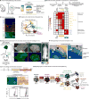
Figure 4
Figure 4. Dorsal-ventral patterning of somite precursors into sclerotome and dermomyotome and downstream progeny
A. Somite patterning in vivo B. qPCR heatmap of hESCs (D0), early somite progenitors (D3) or those differentiated into sclerotome (D4, D5 or D6, using 21K+C59) or dermomyotome (D4 or D5, using BMP4+CHIR+Vismodegib) C. SOX9 and TWIST1 staining of day 6 H7-derived sclerotome; scale bar = 100 μm D. PCA of scRNA-seq from indicated populations; each dot depicts a single cell E. EF1A-BCL2-2A-GFP expressing H9-derived sclerotome was subcutaneously injected into NSG mice; 2 months later, ectopic GFP+ human bones formed (left); bioluminescent imaging of mice 1 month post-transplantation by UBC-Luciferase-2A-tdTomato H9-derived sclerotome F. Russell-Movat’s Pentachrome staining of 2-month-old sclerotome grafts revealed zones of chondrogenesis and ossification, with cartilage stained blue; black line denotes the edge of the graft; white line denotes boundary of the ossifying region; scale bar = 1 mm (bottom) G. COL2A1 (top left) and Safranin-O staining (top right) of D6+2 or D6+6 hESC-derived cartilage, respectively; scale bars = 0.1 mm (left) and 1 mm (right); SMAα intracellular FACS of hESCs or D8 fibroblast-like cells (bottom) H. Somite patterning into dermomyotome or sclerotome and downstream differentiation See also Fig. S4, Fig. S5
Figure 5
Figure 5. Lateral mesoderm patterning into cardiac vs. limb mesoderm fates
A. Cardiac vs. forelimb bifurcation B. To assess the role of WNT in lateral mesoderm patterning, D1 PS was differentiated to lateral mesoderm (30 ng/mL BMP4 + 1 μM C59 + 2 μM SB505124) for varying lengths of time (until D2, D2.5 or D3) and for the last 12 hrs was treated with C59 or 3 μM CHIR (in addition to BS) and qPCR was conducted C. To assess the role of FGF in lateral mesoderm patterning, day 2 NKX2.5-GFP lateral mesoderm was treated with BMP4 + C59 + SB505124 with or without FGF2 (20 ng/mL) or FGFR inhibitor PD173074 (100 nM) for 24 hrs and FACS was conducted on day 3 D. Timepoint FACS of NKX2.5-GFP hESC (Elliott et al., 2011) differentiation using cardiac mesoderm protocol E. Comparison of NKX2.5-GFP+ cell percentages (determined by FACS) on days of differentiation, using the current protocol or a previous method (Burridge et al., 2014) F. Intracellular TNNT2 FACS of H7-derived cardiomyocytes (bottom) G. Electrocardiogram of human fetal heart implanted in the mouse ear, >1 month post-implantation H. 2.5 months post-transplant of EF1A-BCL2-2A-GFP;UBC-tdTomato-Luciferase H9 hESC-derived cardiac lineages into human fetal heart grafts, luciferase+ donor cells were detected (i); engrafted hESC-derived cardiomyocytes were TROPONIN+ and CONNEXIN 43+, scale bar = 40 μm (ii) See also Fig. S6
Figure 6
Figure 6. High-throughput screen for lineage-specific mesoderm surface markers
A. Clustered heatmap of surface marker expression in hESCs and 6 mesoderm derivatives. Each row represents an individual surface marker and color denotes the percentage of cells positive for a given marker. For PS and cardiac mesoderm, marker expression was analyzed after pre-gating on MIXL1-GFP+ and NKX2.5-GFP+ fractions, respectively. B. GARP and DLL1 FACS in hESCs, Day 2 paraxial mesoderm cultures or Day 3 NKX2.5-GFP+ pre-gated cardiac mesoderm C. lrrc32 in situ hybridization in 24 hours post fertilization zebrafish embryo (arrows denote heart) D. DLL1 FACS of day 2 paraxial mesoderm culture (left); qPCR of sorted populations (right) E. scRNA-seq of sorted DLL1+ human paraxial mesoderm; each dot is a single cell F. PDGFRα FACS of day 5 sclerotome population (left); qPCR of sorted PDGFRα+ and PDGFRα populations (center); in situ hybridization for pdgfra expression (right) in 22 hpf zebrafish embryo (arrowheads denote ventral staining in sclerotome) G. scRNA-seq of sorted PDGFRα+ human sclerotome; each dot is a single cell See also Fig. S7, Table S2
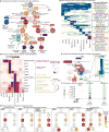
Figure 7
Figure 7. The landscape of mesoderm development
A. Lineage steps with circled numbers correspond to respective sections in the main text and Fig. 1a B. RNA-seq expression of human congenital scoliosis genes C. RNA-seq profiling; color intensity depicts gene expression (log2 TPM) normalized to the expression of that gene in all populations profiled, with the highest-expressing lineage assigned the most intense color value D. ATAC-seq heatmap; each horizontal line depicts a single chromatin element (left, non-binarized in Fig. S8b), with motifs representative of the top 4 lineage-enriched motifs shown (right) E. Inferred trans-regulatory lineages programs (left); heatmap of the 4 FOX TFs most highly expressed in hESC-derived somites (RNA-seq; right) F. ATAC-seq of the MEOX1 locus, with FOX motifs centered in two somitic enhancer elements shown See also Fig. S8

Comment in

References

    1. Ardehali R, Ali SR, Inlay MA, Abilez OJ, Chen MQ, Blauwkamp TA, Yazawa M, Gong Y, Nusse R, Drukker M, et al. Prospective isolation of human embryonic stem cell-derived cardiovascular progenitors that integrate into human fetal heart tissue. Proc Natl Acad Sci USA 2013 - PMC - PubMed
    1. Aulehla A, Wiegraebe W, Baubet V, Wahl MB, Deng C, Taketo M, Lewandoski M, Pourquié O. A β-catenin gradient links the clock and wavefront systems in mouse embryo segmentation. Nature Cell Biology. 2008;10:186–193. - PMC - PubMed
    1. Burridge PW, Matsa E, Shukla P, Lin ZC, Churko JM, Ebert AD, Lan F, Diecke S, Huber B, Mordwinkin NM, et al. Chemically defined generation of human cardiomyocytes. Nature Methods 2014 - PMC - PubMed
    1. Cheung C, Bernardo AS, Trotter MWB, Pedersen RA, Sinha S. Generation of human vascular smooth muscle subtypes provides insight into embryological origin-dependent disease susceptibility. Nat Biotechnol 2012 - PMC - PubMed
    1. Chong JJH, Yang X, Don CW, Minami E, Liu YW, Weyers JJ, Mahoney WM, Van Biber B, Cook SM, Palpant NJ, et al. Human embryonic-stem-cell-derived cardiomyocytes regenerate non-human primate hearts. Nature. 2014;510:273–277. - PMC - PubMed

Publication types

MeSH terms