Randomized Controlled Trial
. 2016 Oct;43(11):2061-71.
doi: 10.1007/s00259-016-3440-3. Epub 2016 Jul 15.

A PET/CT approach to spinal cord metabolism in amyotrophic lateral sclerosis

Affiliations
Randomized Controlled Trial

A PET/CT approach to spinal cord metabolism in amyotrophic lateral sclerosis

Cecilia Marini et al. Eur J Nucl Med Mol Imaging. 2016 Oct.

Abstract

Purpose: In amyotrophic lateral sclerosis, functional alterations within the brain have been intensively assessed, while progression of lower motor neuron damage has scarcely been defined. The aim of the present study was to develop a computational method to systematically evaluate spinal cord metabolism as a tool to monitor disease mechanisms.

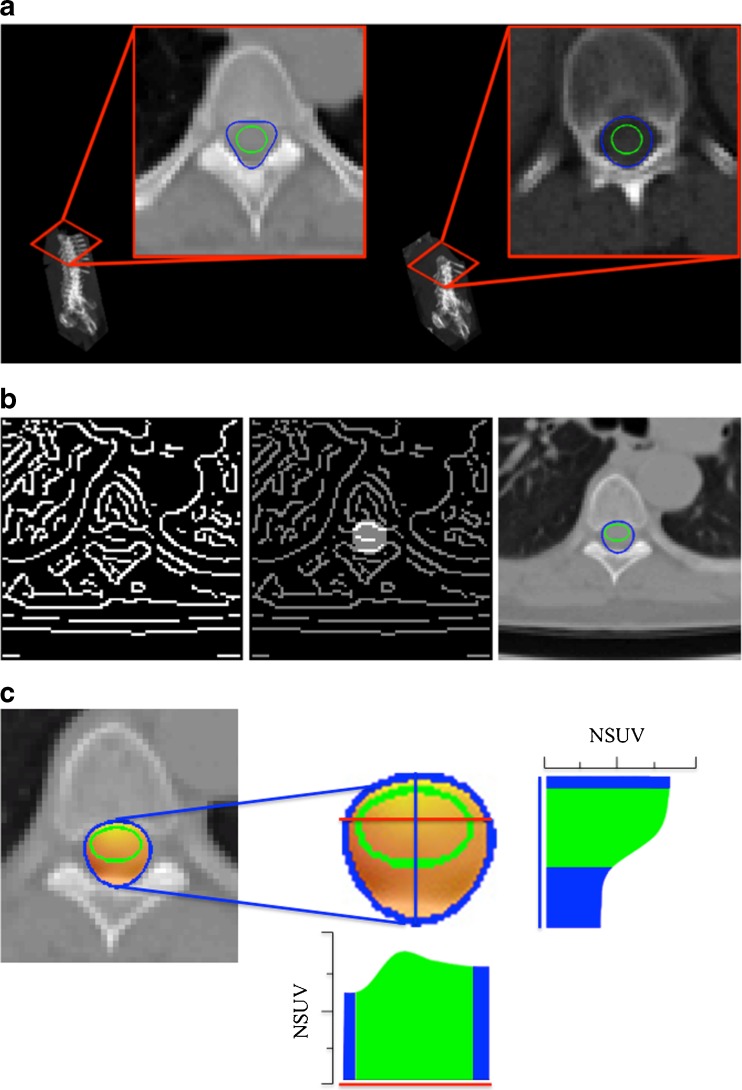
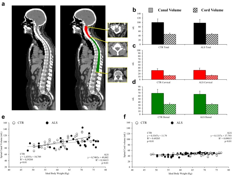
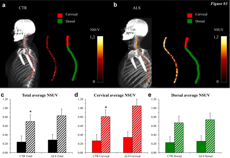
Methods: A new computational three-dimensional method to extract the spinal cord from (18)F-FDG PET/CT images was evaluated in 30 patients with spinal onset amyotrophic lateral sclerosis and 30 controls. The algorithm identified the skeleton on the CT images by using an extension of the Hough transform and then extracted the spinal canal and the spinal cord. In these regions, (18)F-FDG standardized uptake values were measured to estimate the metabolic activity of the spinal canal and cord. Measurements were performed in the cervical and dorsal spine and normalized to the corresponding value in the liver.

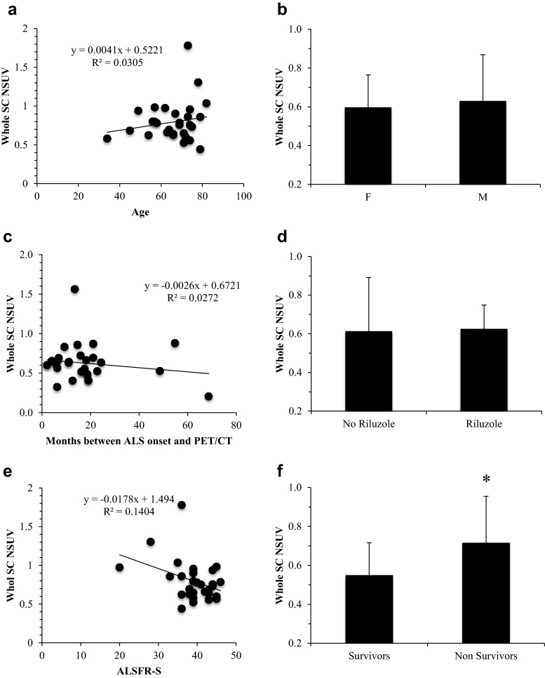
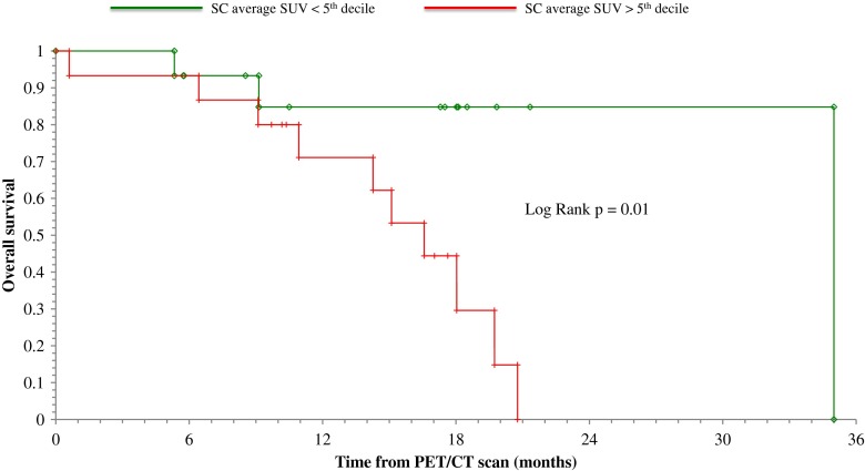
Results: Uptake of (18)F-FDG in the spinal cord was significantly higher in patients than in controls (p < 0.05). By contrast, no significant differences were observed in spinal cord and spinal canal volumes between the two groups. (18)F-FDG uptake was completely independent of age, gender, degree of functional impairment, disease duration and riluzole treatment. Kaplan-Meier analysis showed a higher mortality rate in patients with standardized uptake values above the fifth decile at the 3-year follow-up evaluation (log-rank test, p < 0.01). The independence of this value was confirmed by multivariate Cox analysis.

Conclusion: Our computational three-dimensional method enabled the evaluation of spinal cord metabolism and volume and might represent a potential new window onto the pathophysiology of amyotrophic lateral sclerosis.

Keywords: Amyotrophic lateral sclerosis; Neuroimaging; PET/CT; Spinal cord.


Conflict of interest statement

Compliance with ethical standardsConflicts of interestNone.Ethical approvalAll procedures performed were in accordance with the ethical standards of the institutional and national research committees and with the principles of the 1964 Declaration of Helsinki and its later amendments.Informed consentAll patients provided signed informed consent to be entered into the study that was approved by the Ethics Committees of IRCCS AOU San Martino-IST in Genova and of AUO Città della Salute e della Scienza in Torino, Italy.

Figures

Fig. 1
Fig. 1
a Detection of spinal canal and spinal cord at different heights of the vertebral column. The Hough transform-based procedure with respect to the curve with three convexities allows identification of the spinal canal (blue line), while the spinal cord is detected by the Hough transform-based procedure with respect to the ellipse (green line). b Left to right: edge detection of a CT slice in the vertebral region; edge points inside the region bounded by the curve with three convexities; and the curve with three convexities (blue line) and the ellipse (green line) detected by applying the Hough transform-based procedure. c Left to right: fusion of CT and PET data for the slice considered in b zoomed on the spinal region, and histograms of the behaviour of the normalized standardized uptake values for the same slice and along two specific projections highlighted in red
Fig. 2
Fig. 2
a Sagittal whole-body CT scan (left) and the corresponding image showing extraction of the cervical (red) and dorsal (green) spinal canal. b–d Volumes of the spinal canal (solid bars) and spinal cord (hatched bars) in the control subjects (CTR) and in the 30 ALS patients (ALS): b total volumes, c cervical region, d dorsal region. There are no differences between the control subjects and ALS patients. e, f Linear regression analysis of the relationship between spinal canal volume (e) and spinal cord volume (f) and ideal body weight
Fig. 3
Fig. 3
a, b Whole-body maximum intensity projection CT images coregistered with the extracted PET data for the spinal cord in a control subject (a CTR) and an ALS patient (b ALS). The PET data alone are also shown for the corresponding SCs as the average SUV normalized to the corresponding liver values (average NSUV) together with images of the cervical and dorsal segments. c–e Average NSUV for the spinal canal (solid bars) and spinal cord (hatched bars) in control subjects (CTR) and 30 ALS patients (ALS): c whole spinal cord/spinal canal, d cervical segment, e dorsal segment. FDG uptake in the spinal cord was significantly lower in control subjects for the whole spinal cord (c) and for the cervical segment (d), but not for the dorsal segment (e). *p < 0.05
Fig. 4
Fig. 4
a, c, e Linear regression analysis of the relationship between spinal cord (SC) NSUV and patient age (a), time between diagnosis and imaging (c) and revised ALS functional rating scale (ALSFR-S) score (e). b, d, f The spinal cord metabolic pattern (in terms of NSUV) was not significantly different between male (M) and female (F) patients (b), or between those treated and those not treated with riluzole (d), but SC NSUV was significantly higher in patients who had died (Nonsurvivors) than in those who were still alive (Survivors) at the end of the 36-month follow-up period (f). *p < 0.05
Fig. 5
Fig. 5
Kaplan-Meier curves showing overall survival in patients with SC_NSUV above and below the fifth decile. High FDG uptake in the whole spinal cord was associated with a higher mortality rate

References

    1. Rowland LP, Shneider NA. Amyotrophic lateral sclerosis. N Engl J Med. 2001;344:1688–1700. doi: 10.1056/NEJM200105313442207. - DOI - PubMed
    1. Chiò A, Pagani M, Agosta F, Calvo A, Cistaro A, Filippi M. Neuroimaging in amyotrophic lateral sclerosis: insights into structural and functional changes. Lancet Neurol. 2014;13:1228–1240. doi: 10.1016/S1474-4422(14)70167-X. - DOI - PubMed
    1. Pagani M, Chiò A, Valentini MC, Öberg J, Nobili F, Calvo A, et al. Functional pattern of brain FDG-PET in amyotrophic lateral sclerosis. Neurology. 2014;83:1067–1074. doi: 10.1212/WNL.0000000000000792. - DOI - PubMed
    1. Cistaro A, Pagani M, Montuschi A, Calvo A, Moglia C, Canosa A, et al. The metabolic signature of C9ORF72-related ALS: FDG PET comparison with nonmutated patients. Eur J Nucl Med Mol Imaging. 2014;41:844–852. doi: 10.1007/s00259-013-2667-5. - DOI - PMC - PubMed
    1. Cistaro A, Valentini MC, Chiò A, Nobili F, Calvo A, Moglia C, et al. Brain hypermetabolism in amyotrophic lateral sclerosis: a FDG PET study in ALS of spinal and bulbar onset. Eur J Nucl Med Mol Imaging. 2012;39:251–259. doi: 10.1007/s00259-011-1979-6. - DOI - PubMed

Publication types

MeSH terms

Substances