. 2016 Jul 31;428(15):2997-3012.
doi: 10.1016/j.jmb.2016.07.002. Epub 2016 Jul 14.

Processive DNA Unwinding by RecBCD Helicase in the Absence of Canonical Motor Translocation

Affiliations

Processive DNA Unwinding by RecBCD Helicase in the Absence of Canonical Motor Translocation

Michael J Simon et al. J Mol Biol. .

Abstract

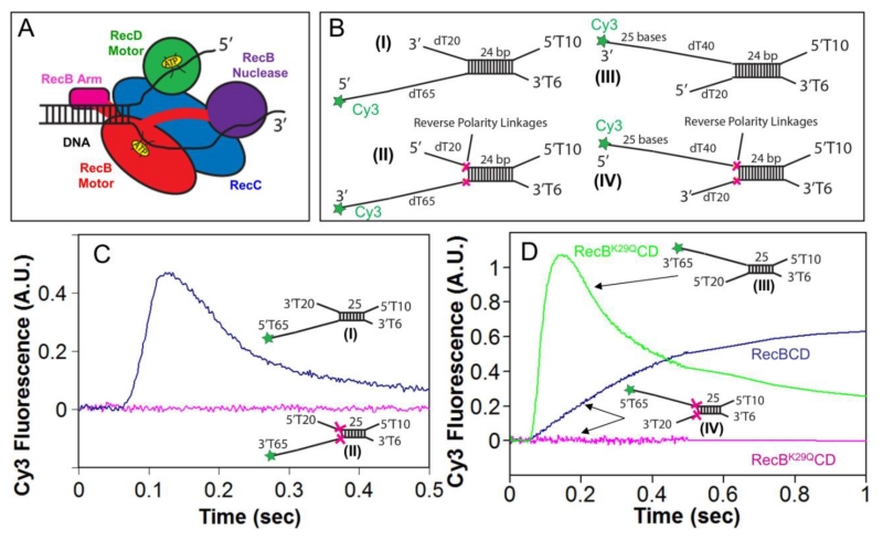
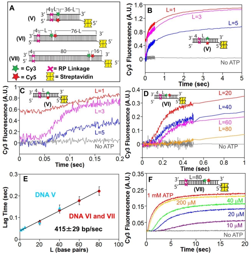
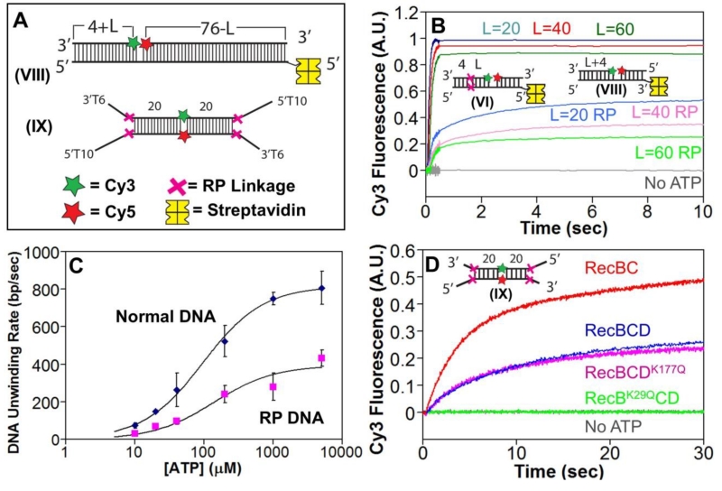
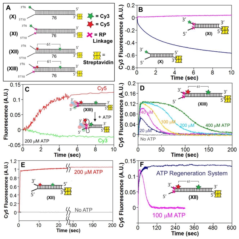
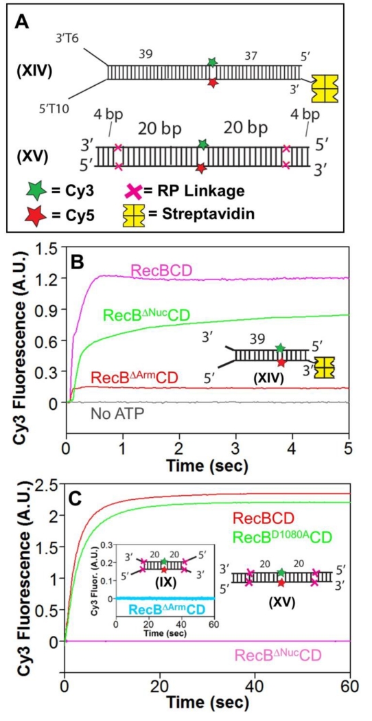
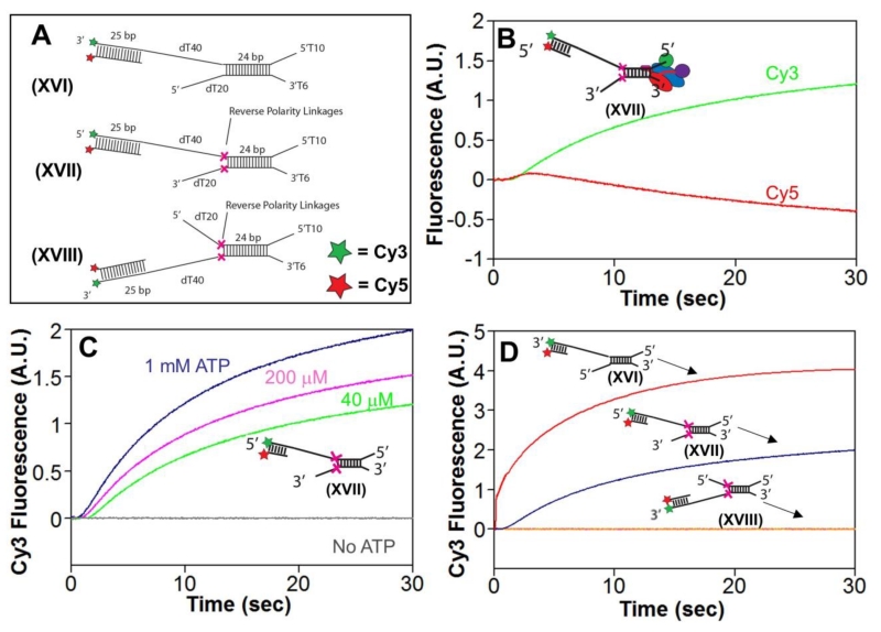
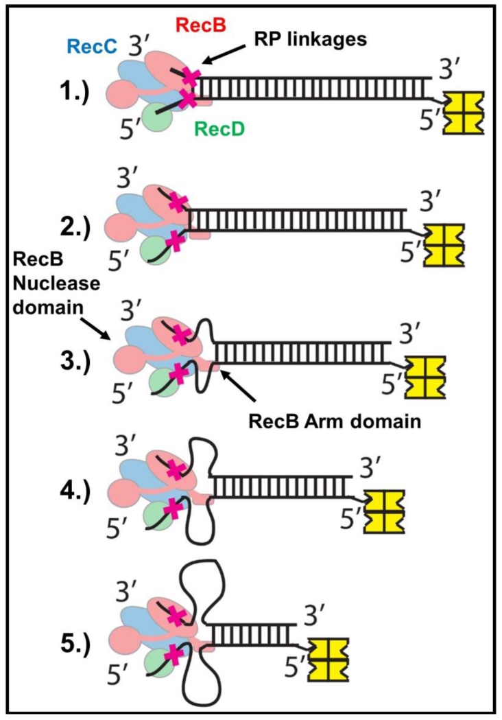
Escherichia coli RecBCD is a DNA helicase/nuclease that functions in double-stranded DNA break repair. RecBCD possesses two motors (RecB, a 3' to 5' translocase, and RecD, a 5' to 3' translocase). Current DNA unwinding models propose that motor translocation is tightly coupled to base pair melting. However, some biochemical evidence suggests that DNA melting of multiple base pairs may occur separately from single-stranded DNA translocation. To test this hypothesis, we designed DNA substrates containing reverse backbone polarity linkages that prevent ssDNA translocation of the canonical RecB and RecD motors. Surprisingly, we find that RecBCD can processively unwind DNA for at least 80bp beyond the reverse polarity linkages. This ability requires an ATPase active RecB motor, the RecB "arm" domain, and also the RecB nuclease domain, but not its nuclease activity. These results indicate that RecBCD can unwind duplex DNA processively in the absence of ssDNA translocation by the canonical motors and that the nuclease domain regulates the helicase activity of RecBCD.

Keywords: SF1 helicase; allostery; fluorescence; recombination.


Figures

Figure 1
Figure 1. Reversal of the sugar-phosphate backbone polarity prevents ssDNA translocation of the RecB and RecD canonical motors
A. Cartoon of RecBCD bound to a dsDNA end based on a crystal structure[3]. RecB, RecC, and RecD are shown in red, blue, and green, respectively, with the RecB arm and nuclease domains shown in pink and purple. B. DNA substrates used to test the effects of reverse polarity (RP) linkages on ssDNA translocation. The red X indicates the positions of the 3′-3′ and 5′-5′ RP linkages. A green star indicates the position of a Cy3 fluorophore. C. Stopped-flow time courses monitoring Cy3 fluorescence were obtained by pre-incubating RecBCD (18.75 nM) with DNA I (blue trace) or DNA II (red trace) (25 nM) in Buffer M (250 mM NaCl) with ATP (1 mM) and heparin (8 mg/mL) in buffer M (8 mM NaCl) at 1:10 volumetric ratio yielding a final NaCl concentration of 30 mM (concentrations of RecBCD and DNA listed are after mixing) at 25° C. D. Stopped-flow time courses monitoring Cy3 fluorescence performed as described for panel C. RecBK29QCD pre-incubated with DNA III (green trace) or DNA IV (red trace); RecBCD pre-incubated with DNA IV (blue trace).
Figure 2
Figure 2. RecBCD can unwind DNA when canonical motor translocation is blocked by RP linkages
A. DNA used to test if RecBCD can unwind DNA when ssDNA translocation is blocked by RP linkages. A red X indicates the positions of the 3′-3′ and 5′-5′ RP linkages, separated by L bp from a Cy3 (green star) and Cy5 (red star) fluorophores. One end of each DNA is biotinylated so that streptavidin (yellow) can bind and block binding of RecBCD. B. Stopped-flow time courses were obtained by mixing pre-incubated RecBCD (15 nM) and DNA V (20 nM) with ATP (5 mM) and heparin (8 mg/mL) in Buffer M (30 mM NaCl) at 25° C. Cy3 fluorescence was excited at 505 nm and Cy3 and Cy5 fluorescence were monitored simultaneously. Cy3 fluorescence time courses are shown for DNA V with L=1 bp (red), L=3 bp (pink) and L=5 bp (blue). C. The first 0.2 seconds of the time courses shown in panel B showing increase in lag time with increasing L. D. Stopped-flow time courses showing RecBCD unwinding of DNA VI and DNA VII. Experiments were performed as described in panel B. Cy3 time courses for DNA series VI with L=20 bp (red), L=40 bp (blue), L=60 bp (pink) and DNA VII (L=80 bp). E. Lag times for RecBCD unwinding of RP DNA (5 mM ATP) with Cy3/Cy5 positioned in the DNA backbone (DNA series V) (blue triangles) or across a nick (DNA VI and VII) (red). F. ATP-dependence of RecBCD unwinding of DNA VII. Experiments were performed as described in panel B, at the indicated [ATP].
Figure 3
Figure 3. RecBCD unwinds RP DNA with a two-fold slower rate and requires an active RecB motor
A. DNA VIII used to monitor unwinding of normal DNA. Each end of DNA IX contains a high affinity site for RecBCD binding. A red X indicates the positions of the 3′-3′ and 5′-5′ RP linkages, separated by L bp from a Cy3 (green star) and Cy5 (red star) fluorophores. DNA VIII is biotinylated at one end so that streptavidin (yellow) can bind and block binding of RecBCD. B. Comparison of RecBCD unwinding time courses with normal DNA (series VIII) vs. RP DNA (series VI) performed by mixing pre-incubated RecBCD (15 nM) and DNA (20 nM) with ATP (5 mM) and heparin (8 mg/mL) in Buffer M (30 mM NaCl) at 25° C. Cy3 fluorescence was excited at 505 nm, and Cy3 and Cy5 fluorescence were monitored simultaneously. Cy3 fluorescence time courses for DNA with L=20 bp (blue), L=40 bp (red), and L=60 bp (green) are shown for normal DNA Series VIII and RP DNA Series VI. C. . ATP dependence of DNA unwinding rates for normal DNA (blue) and RP DNA (pink). Smooth curves are the best fits to the Michaelis-Menten equation yielding Km=96±8 μM, Vmax=815±16 bp/sec for normal DNA and Km=138±57, Vmax=392±38 for RP DNA. D. Stopped flow time courses, monitoring Cy3 fluorescence, for RecBCD (blue), RecBC (red), RecBCDK177Q (pink), and RecBK29QCD (green) with DNA IX.
Figure 4
Figure 4. The RecBCD motors remain stuck behind the RP linkages and induce ssDNA loops during DNA unwinding
A. DNA substrates used to test for ssDNA loop formation during RecBCD unwinding of RP DNA. A red X indicates the positions of the 3′-3′ and 5′-5′ RP linkages. Cy3 (green star) and Cy5 (red star) fluorophores are separated by 61 base pairs. One end of each DNA is biotinylated so that streptavidin (yellow) can bind and block binding of RecBCD. B. Stopped-flow time courses, monitoring Cy3 fluorescence, for RecBCD unwinding of normal DNA X (blue) and RP DNA XI (pink). RecBCD (18.75 nM) and DNA (25 nM) were pre-incubated and then mixed with ATP (5 mM) and heparin (8 mg/mL) in Buffer M (30 mM NaCl) at 25° C. C. Stopped-flow time courses showing an increase in FRET signal accompanying RecBD1080ACD unwinding of RP DNA XIII. Experiments were performed by mixing pre-incubated RecBCD (37.5 nM) and DNA XIII (50 nM) with ATP (200 μM) and heparin (8 mg/mL) in Buffer M (30 mM NaCl) at 25° C. Cy3 fluorescence was excited at 505 nm, and Cy3 (green) and Cy5 (red) fluorescence were monitored. D. Cy3 fluorescence time courses for experiments performed with RecBD1080ACD and RP DNA XIII as described in panel C, at 400 μM ATP (green), 200 μM ATP (light blue), 100 μM ATP (gold), 40 μM ATP (pink), 20 μM ATP (dark blue), and no ATP (gray). Inset-identical stopped-flow experiment performed with RecBD1080ACD and DNA XII at 200 μM ATP.
Figure 5
Figure 5. The RecB arm and nuclease domains are needed for RecBCD to unwind RP DNA
A. DNA substrates used to test for unwinding by RecBΔArmCD and RecBΔNucCD. DNA XIV contains a high affinity binding site on one end and is biotinylated on the opposite end to bind streptavidin (yellow) to block RecBCD binding. Cy3 (green star) and Cy5 (red star) are on opposite strands 39 bp from the binding site. A red X indicates the positions of the 3′-3′ and 5′-5′ RP linkages in DNA XV separated by L bp from a Cy3 (green star) and Cy5 (red star) fluorophores. B. Stopped-flow Cy3 fluorescence time courses showing unwinding of DNA XIV (20 nM) by RecBCD, RecBΔNucCD, and RecBΔArmCD (15 nM) in Buffer M (30 mM NaCl) (5 mM ATP and 8 mM heparin) at 25° C. C. Stopped-flow Cy3 fluorescence time courses with RP DNA XV and RecBCD (15 nM) (red), RecBD1080ACD (15 nM) (green), and RecBΔNucCD (15 nM) (pink) in Buffer M30 (5 mM ATP and 8 mg/mL heparin) at 25° C. Inset-RecBΔArmCD is unable to unwind RP DNA IX containing a high affinity binding site under the same conditions.
Figure 6
Figure 6. RecBCD unwinding of RP DNA requires its secondary translocase activity
A. Substrates used to test re-initiation of unwinding by RecBCD. B. Stopped-flow experiments were performed by mixing pre-incubated RecBCD and DNA Substrate XVII (18.75 and 25 nM, respectively) in Buffer M (250 mM NaCl) in a 1:10 volumetric ratio with ATP (1 mM) and heparin (8 mg/mL) in buffer M (8 mM NaCl) yielding the final concentrations listed above and a final NaCl concentration of 30 mM at 25°C. Cy3 fluorescence was excited at 505 nm, and Cy3 fluorescence (green) was monitored at 570 nm and Cy5 fluorescence (red) was monitored at > 665 nm. C. Cy3 fluorescence traces for experiments as performed in panel B, at 1 mM ATP (dark blue), 200 μM ATP (pink), 40 μM ATP (light blue), and no ATP (gray). D. Cy3 fluorescence traces for experiments as performed in panel B at 1 mM ATP using Substrate XVI (red), XVII (blue), or XVIII (orange).
Figure 7
Figure 7. Proposed mechanism for unwinding of reversed polarity DNA by RecBCD
(Step 1) RecBCD binds to and melts 6 base pairs at a normal DNA end. (Step 2) Canonical ssDNA motors (RecB (red) and RecD (green)) translocate along the ssDNA tracks until the RP linkages (red X) are encountered. (Step 3-5) RP linkages block ssDNA translocation by the canonical RecB and RecD motors, but the secondary translocase, fueled by ATP hydrolysis in the RecB motor, simultaneously pulls dsDNA toward RecBCD, resulting in the formation of single stranded DNA loops. The RecB Arm and RecB Nuclease domains (highlighted) are necessary for this activity.

Similar articles

Cited by

References

    1. Dillingham MS, Kowalczykowski SC. RecBCD Enzyme and the Repair of Double-Stranded DNA Breaks. Microbiology and molecular biology reviews : MMBR. 2008;72:642–71. - PMC - PubMed
    1. Smith GR. How RecBCD enzyme and Chi promote DNA break repair and recombination: a molecular biologist’s view. Microbiology and molecular biology reviews : MMBR. 2012;76:217–28. - PMC - PubMed
    1. Singleton MR, Dillingham MS, Gaudier M, Kowalczykowski SC, Wigley DB. Crystal structure of RecBCD enzyme reveals a machine for processing DNA breaks. Nature. 2004;432:187–93. - PubMed
    1. Chen HW, Ruan B, Yu M, Wang J, Julin DA. The RecD subunit of the RecBCD enzyme from Escherichia coli is a single-stranded DNA-dependent ATPase. J Biol Chem. 1997;272:10072–9. - PubMed
    1. Taylor AF, Smith GR. RecBCD enzyme is a DNA helicase with fast and slow motors of opposite polarity. Nature. 2003;423:889–93. - PubMed

MeSH terms

LinkOut - more resources