

Predicting Malignant Nodules from Screening CT Scans
- PMID: 27422797
- PMCID: PMC5545995
- DOI: 10.1016/j.jtho.2016.07.002
Predicting Malignant Nodules from Screening CT Scans
Erratum in
-
Erratum.J Thorac Oncol. 2018 Feb;13(2):280-281. doi: 10.1016/j.jtho.2017.09.1959. J Thorac Oncol. 2018. PMID: 29425613 No abstract available.
Abstract
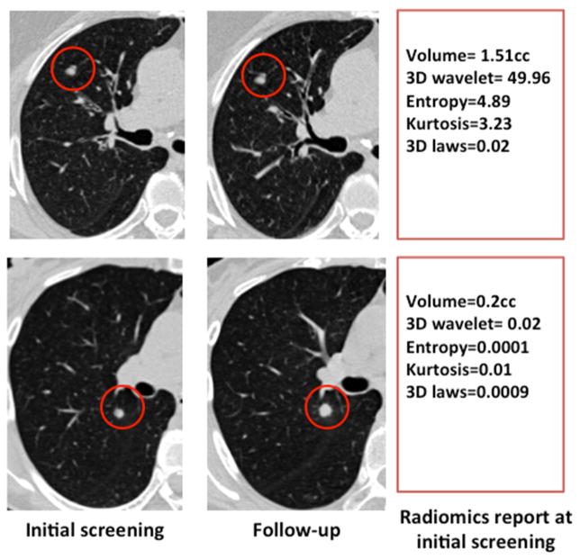
Objectives: The aim of this study was to determine whether quantitative analyses ("radiomics") of low-dose computed tomography lung cancer screening images at baseline can predict subsequent emergence of cancer.
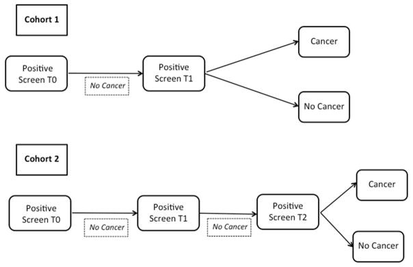
Methods: Public data from the National Lung Screening Trial (ACRIN 6684) were assembled into two cohorts of 104 and 92 patients with screen-detected lung cancer and then matched with cohorts of 208 and 196 screening subjects with benign pulmonary nodules. Image features were extracted from each nodule and used to predict the subsequent emergence of cancer.
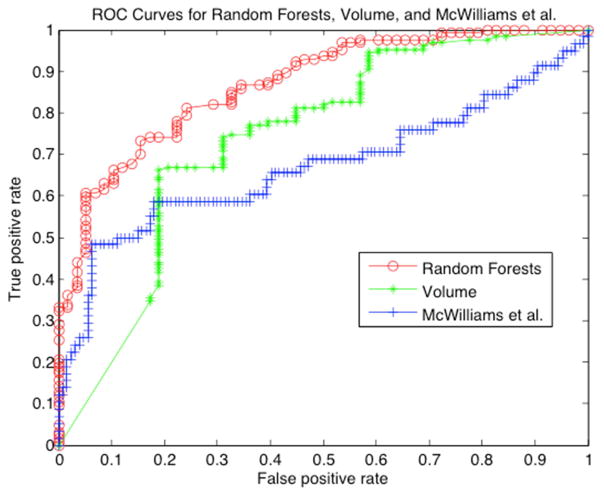
Results: The best models used 23 stable features in a random forests classifier and could predict nodules that would become cancerous 1 and 2 years hence with accuracies of 80% (area under the curve 0.83) and 79% (area under the curve 0.75), respectively. Radiomics outperformed the Lung Imaging Reporting and Data System and volume-only approaches. The performance of the McWilliams risk assessment model was commensurate.
Conclusions: The radiomics of lung cancer screening computed tomography scans at baseline can be used to assess risk for development of cancer.
Keywords: Computed tomography; Lung cancer; Machine learning; Prediction; Radiomics; Screening.
Copyright © 2016 International Association for the Study of Lung Cancer. Published by Elsevier Inc. All rights reserved.
Figures
Comment in
-
Predicting Malignant Nodules from Screening CTs.J Thorac Oncol. 2016 Dec;11(12):2045-2047. doi: 10.1016/j.jtho.2016.09.117. J Thorac Oncol. 2016. PMID: 27866632 No abstract available.
Similar articles
-
Preoperative diagnosis of malignant pulmonary nodules in lung cancer screening with a radiomics nomogram.Cancer Commun (Lond). 2020 Jan;40(1):16-24. doi: 10.1002/cac2.12002. Epub 2020 Mar 3. Cancer Commun (Lond). 2020. PMID: 32125097 Free PMC article.
-
Leveraging Serial Low-Dose CT Scans in Radiomics-based Reinforcement Learning to Improve Early Diagnosis of Lung Cancer at Baseline Screening.Radiol Cardiothorac Imaging. 2024 Jun;6(3):e230196. doi: 10.1148/ryct.230196. Radiol Cardiothorac Imaging. 2024. PMID: 38752718 Free PMC article.
-
Localized thin-section CT with radiomics feature extraction and machine learning to classify early-detected pulmonary nodules from lung cancer screening.Phys Med Biol. 2018 Mar 14;63(6):065005. doi: 10.1088/1361-6560/aaafab. Phys Med Biol. 2018. PMID: 29446758
-
Interpreting Lung Cancer Screening CTs: Practical Approach to Lung Cancer Screening and Application of Lung-RADS.Clin Chest Med. 2024 Jun;45(2):279-293. doi: 10.1016/j.ccm.2023.08.014. Epub 2023 Sep 15. Clin Chest Med. 2024. PMID: 38816088 Review.
-
Benefits and harms in the National Lung Screening Trial: expected outcomes with a modern management protocol.Lancet Respir Med. 2019 Aug;7(8):655-656. doi: 10.1016/S2213-2600(19)30136-5. Epub 2019 May 7. Lancet Respir Med. 2019. PMID: 31076382 Free PMC article. Review. No abstract available.
Cited by
-
Radiomics Prediction of EGFR Status in Lung Cancer-Our Experience in Using Multiple Feature Extractors and The Cancer Imaging Archive Data.Tomography. 2020 Jun;6(2):223-230. doi: 10.18383/j.tom.2020.00017. Tomography. 2020. PMID: 32548300 Free PMC article.
-
Do we need to see to believe?-radiomics for lung nodule classification and lung cancer risk stratification.J Thorac Dis. 2020 Jun;12(6):3303-3316. doi: 10.21037/jtd.2020.03.105. J Thorac Dis. 2020. PMID: 32642254 Free PMC article. Review.
-
Prediction of Nodal Metastasis in Lung Cancer Using Deep Learning of Endobronchial Ultrasound Images.Cancers (Basel). 2022 Jul 8;14(14):3334. doi: 10.3390/cancers14143334. Cancers (Basel). 2022. PMID: 35884395 Free PMC article.
-
CT-based radiomics signature for the stratification of N2 disease risk in clinical stage I lung adenocarcinoma.Transl Lung Cancer Res. 2019 Dec;8(6):876-885. doi: 10.21037/tlcr.2019.11.18. Transl Lung Cancer Res. 2019. PMID: 32010566 Free PMC article.
-
[A clinical-radiomics nomogram for differentiating focal organizing pneumonia and lung adenocarcinoma].Nan Fang Yi Ke Da Xue Xue Bao. 2024 Feb 20;44(2):397-404. doi: 10.12122/j.issn.1673-4254.2024.02.23. Nan Fang Yi Ke Da Xue Xue Bao. 2024. PMID: 38501426 Free PMC article. Chinese.
References
-
- American Cancer Society. Cancer Facts & Figures 2015. Atlanta: American Cancer Society; 2015.
-
- American Cancer Society. Global Cancer Facts & Figures. 2. Atlanta: American Cancer Society; 2011.
-
- Gopal M, Abdullah SE, Grady JJ, et al. Screening for lung cancer with low-dose computed tomography: a systematic review and meta-analysis of the baseline findings of randomized controlled trials. Journal of thoracic oncology : official publication of the International Association for the Study of Lung Cancer. 2010;5:1233–9. - PMC - PubMed
Publication types
MeSH terms
Grants and funding
LinkOut - more resources
Full Text Sources
Other Literature Sources
Medical
