. 2016 Aug 16;36(4):e00369.
doi: 10.1042/BSR20160075. Print 2016 Aug.

Effects of serum amyloid A on the structure and antioxidant ability of high-density lipoprotein

Affiliations

Effects of serum amyloid A on the structure and antioxidant ability of high-density lipoprotein

Megumi Sato et al. Biosci Rep. .

Abstract

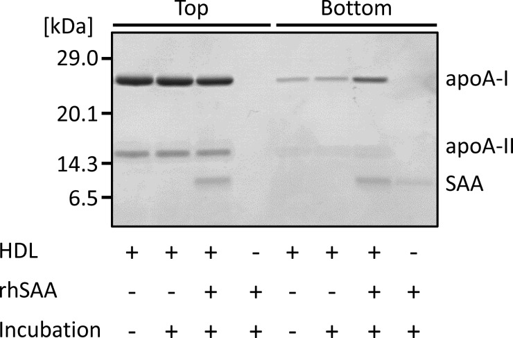
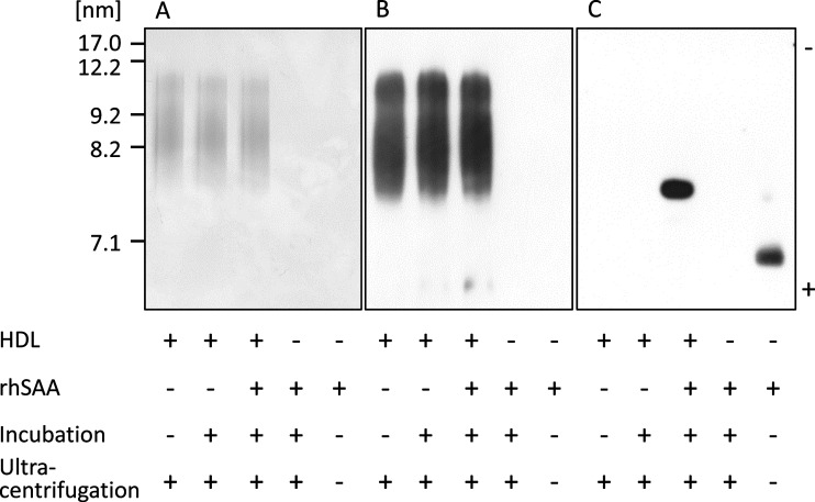
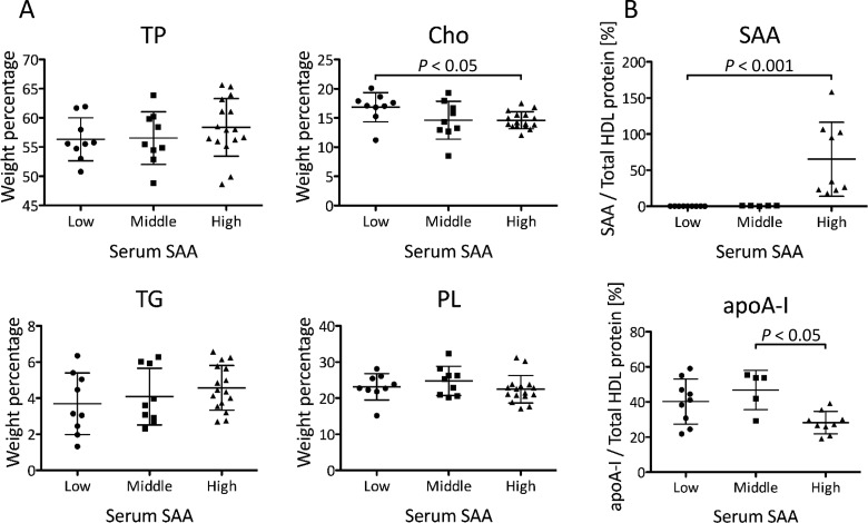
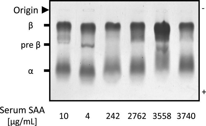
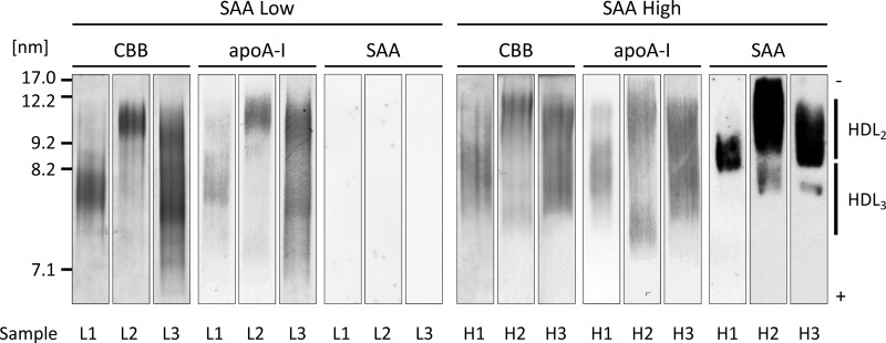
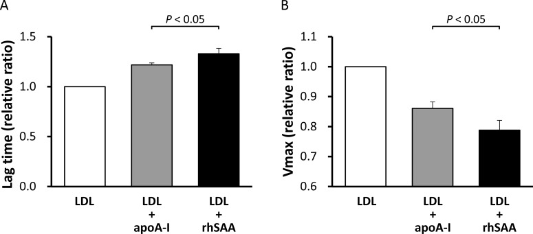
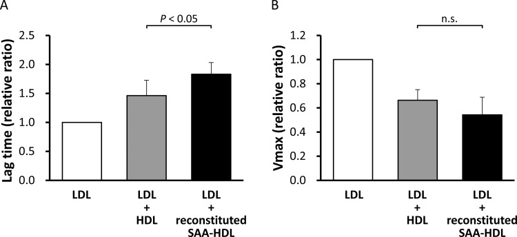
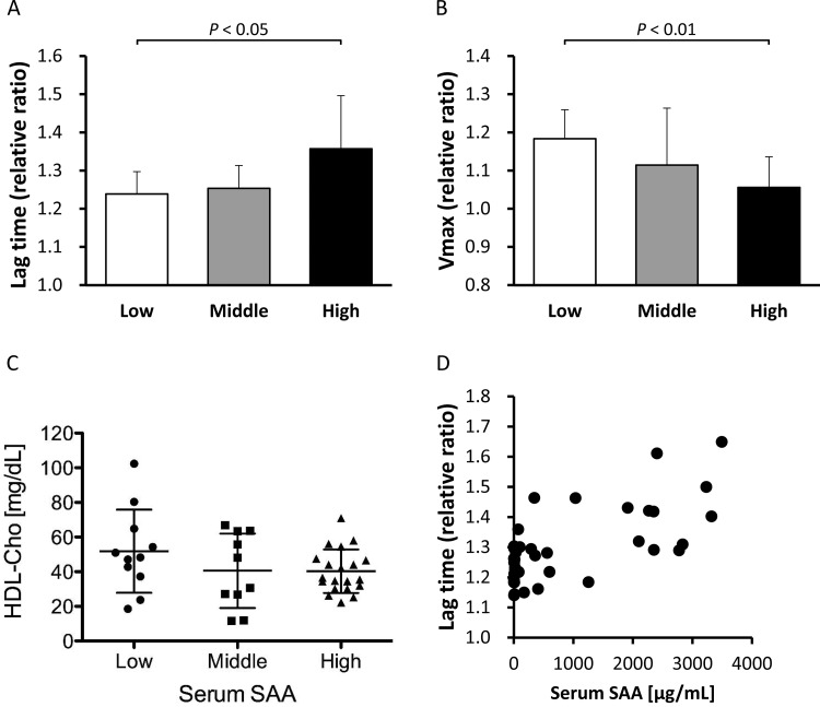
Serum amyloid A (SAA) levels increase during acute and chronic inflammation and are mainly associated with high-density lipoprotein (HDL). In the present study, we investigated the effect of SAA on the composition, surface charge, particle size and antioxidant ability of HDL using recombinant human SAA (rhSAA) and HDL samples from patients with inflammation. We confirmed that rhSAA bound to HDL3 and released apolipoprotein A-I (apoA-I) from HDL without an apparent change in particle size. Forty-one patients were stratified into three groups based on serum SAA concentrations: Low (SAA ≤ 8 μg/ml), Middle (8 < SAA ≤ 100 μg/ml) and High (SAA > 100 μg/ml). The ratios of apoA-I to total protein mass, relative cholesterol content and negative charge of HDL samples obtained from patients with high SAA levels were lower than that for samples from patients with low SAA levels. Various particle sizes of HDL were observed in three groups regardless of serum SAA levels. Antioxidant ability of rhSAA, evaluated as the effect on the formation of conjugated diene in low-density lipoprotein (LDL) induced by oxidation using copper sulfate, was higher than that of apoA-I. Consistent with this result, reconstituted SAA-containing HDL (SAA-HDL) indicated higher antioxidant ability compared with normal HDL. Furthermore, HDL samples obtained from High SAA group patients also showed the highest antioxidant ability among the three groups. Consequently, SAA affects the composition and surface charge of HDL by displacement of apoA-I and enhances its antioxidant ability.

Keywords: antioxidant ability; high-density lipoprotein; inflammation; serum amyloid A.


Figures

Figure 1
Figure 1. Displacement of apoA-I by SAA
rhSAA (final concentration of 1 mg/ml) was incubated with HDL obtained from healthy subjects (final HDL-Cho of 25 mg/dl) for 60 min at room temperature. The mixture was ultracentrifuged at the density of 1.210 g/ml. Top and bottom fractions were analysed by SDS/PAGE and visualized by CBB staining. The figure is representative of four independent experiments.
Figure 2
Figure 2. Features of HDL remodelled by SAA
rhSAA (final concentration of 1 mg/ml) was incubated with HDL obtained from healthy subjects (final HDL-Cho of 25 mg/dl) for 60 min at room temperature. The mixture was ultracentrifuged at the density of 1.210 g/ml. Top fraction was analysed by non-denaturing gel electrophoresis and visualized by CBB staining (A) and western blotting using anti-apoA-I antibody (B) and anti-SAA antibody (C). The figure is representative of four independent experiments.
Figure 3
Figure 3. Composition of HDL obtained from the patients
HDL samples were isolated from the patients with low SAA levels (Low, SAA ≤ 8 μg/ml), middle SAA levels (Middle, 8 < SAA ≤ 100 μg/ml) and high SAA levels (High, SAA > 100 μg/ml), and compared in terms of protein and lipid compositions. (A) Weight percentage of each component against total amount of protein, Cho, TG and PL in HDL was indicated (Low; n=9, Middle; n=9, High; n=16). (B) Weight percentage of SAA and apoA-I against total protein in HDL was indicated (Low; n=9, Middle; n=5, High; n=9). The samples with SAA or apoA-I levels lower than the minimum detectable were excluded. Data are shown as the mean ± S.D.
Figure 4
Figure 4. Surface charge of HDL obtained from the patients
Sera from six patients were analysed by agarose gel electrophoresis followed by staining with Fat Red 7B.
Figure 5
Figure 5. Distribution of SAA in HDL obtained from the patients
HDL isolated from three patients from the Low SAA group (L1–3) and High SAA group (H1–3), respectively, were analysed by non-denaturing gel electrophoresis and visualized by CBB staining and by western blotting using anti-apoA-I antibody and anti-SAA antibody. These are representative patterns of HDL obtained from X and Y patients from Low SAA and High SAA groups, respectively. Serum SAA concentrations of the patients L1, L2, L3, H1, H2 and H3 were 6.0, 3.0, 3.0, 557, 3558 and 2762 μg/ml, respectively.
Figure 6
Figure 6. Antioxidant ability of rhSAA and apoA-I
Antioxidant abilities of rhSAA and apoA-I were determined as described in the ‘Materials and Methods’ section. The values were expressed as the mean of the lag time (A) and Vmax (B) relative to those of LDL alone, defined as 1. Bars represent S.D. from triplicate experiments.
Figure 7
Figure 7. Antioxidant ability of reconstituted SAA-HDL and normal HDL
HDLs obtained from healthy subjects (final HDL-Cho of 25 mg/dl) were incubated with or without rhSAA (final concentration of 1 mg/ml) for 60 min at room temperature. The mixtures were then ultracentrifuged at the density of 1.210 g/ml to remove unattached SAA. HDLs in top fractions were defined as reconstituted SAA-HDL (incubated with rhSAA) and normal HDL (incubated without rhSAA). Antioxidant abilities of both HDLs were determined as described in the ‘Materials and Methods’ section. The values were expressed as the mean of lag time (A) and Vmax (B) relative to those of LDL alone, defined as 1. Bars represent S.D. from four separate experiments. n.s., not significant.
Figure 8
Figure 8. Antioxidant ability of HDL obtained from the patients
Antioxidant abilities of HDLs obtained from the patients, stratified into three groups according to the concentration of serum SAA, Low (SAA ≤ 8 μg/ml, n=11), Middle (8 < SAA ≤ 100 μg/ml, n=10) and High (SAA > 100 μg/ml, n=20), were determined as described in the ‘Materials and Methods’ section. The values were expressed as the mean of lag time (A) and Vmax (B) relative to those of LDL alone, defined as 1. Bars represent S.D. HDL-Cho levels in three groups (C) were also compared. Correlation between relative lag time and serum SAA levels (D) was analysed in all samples of three groups.

References

    1. Coetzee G.A., Strachan A.F., van der Westhuyzen D.R., Hoppe H.C., Jeenah M.S., de Beer F.C. Serum amyloid A-containing human high density lipoprotein 3. Density, size, and apolipoprotein composition. J. Biol. Chem. 1986;261:9644–9651. - PubMed
    1. Chait A., Han C.Y., Oram J.F., Heinecke J.W. Thematic review series: the immune system and atherogenesis. Lipoprotein-associated inflammatory proteins: markers or mediators of cardiovascular disease? J. Lipid Res. 2005;46:389–403. doi: 10.1194/jlr.R400017-JLR200. - DOI - PubMed
    1. Westermark G.T., Fändrich M., Westermark P. AA amyloidosis: pathogenesis and targeted therapy. Annu. Rev. Pathol. 2015;10:321–344. doi: 10.1146/annurev-pathol-020712-163913. - DOI - PubMed
    1. van der Westhuyzen D.R., de Beer F.C., Webb N.R. HDL cholesterol transport during inflammation. Curr. Opin. Lipidol. 2007;18:147–151. doi: 10.1097/MOL.0b013e328051b4fe. - DOI - PubMed
    1. Kisilevsky R., Manley P.N. Acute-phase serum amyloid A: perspectives on its physiological and pathological roles. Amyloid. 2012;19:5–14. doi: 10.3109/13506129.2011.654294. - DOI - PubMed

Publication types