. 2016 Oct:9:77-89.
doi: 10.1016/j.redox.2016.06.004. Epub 2016 Jun 27.

A biphasic effect of TNF-α in regulation of the Keap1/Nrf2 pathway in cardiomyocytes

Affiliations

A biphasic effect of TNF-α in regulation of the Keap1/Nrf2 pathway in cardiomyocytes

Gobinath Shanmugam et al. Redox Biol. 2016 Oct.

Abstract

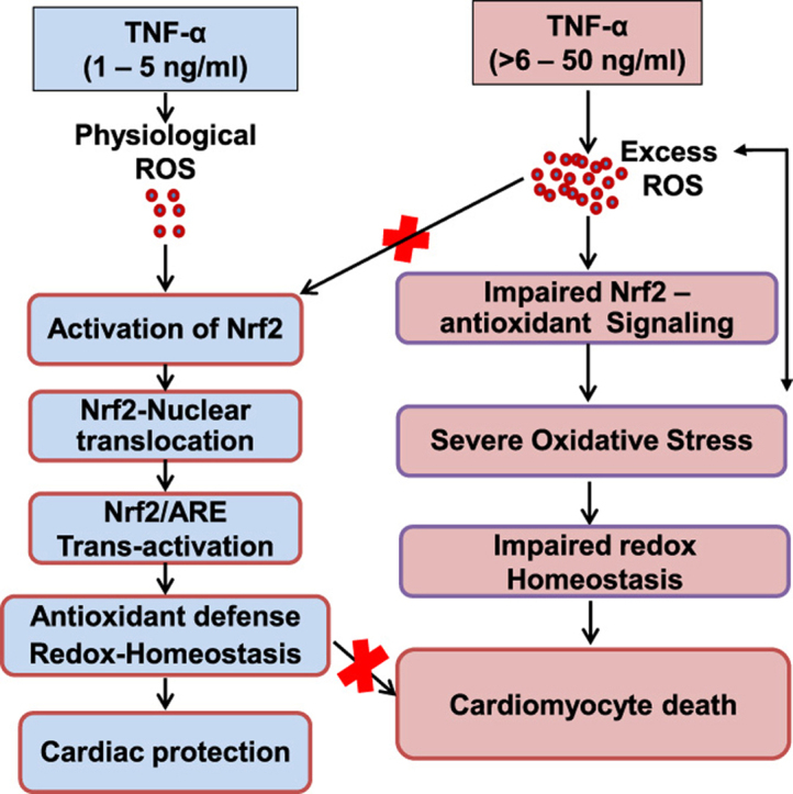
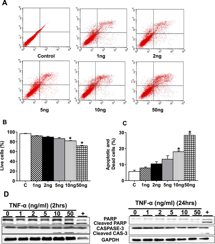
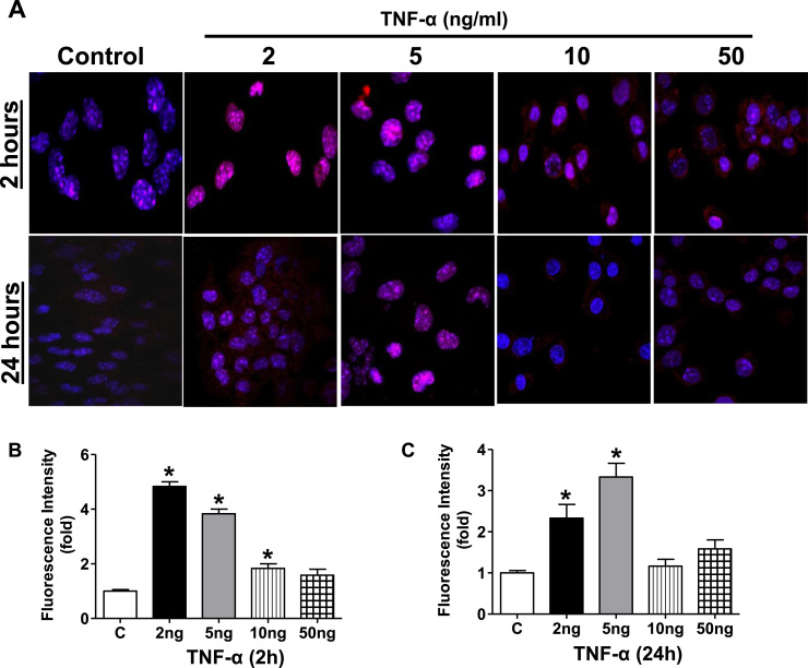
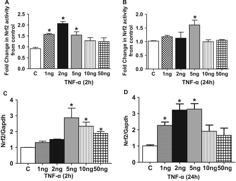
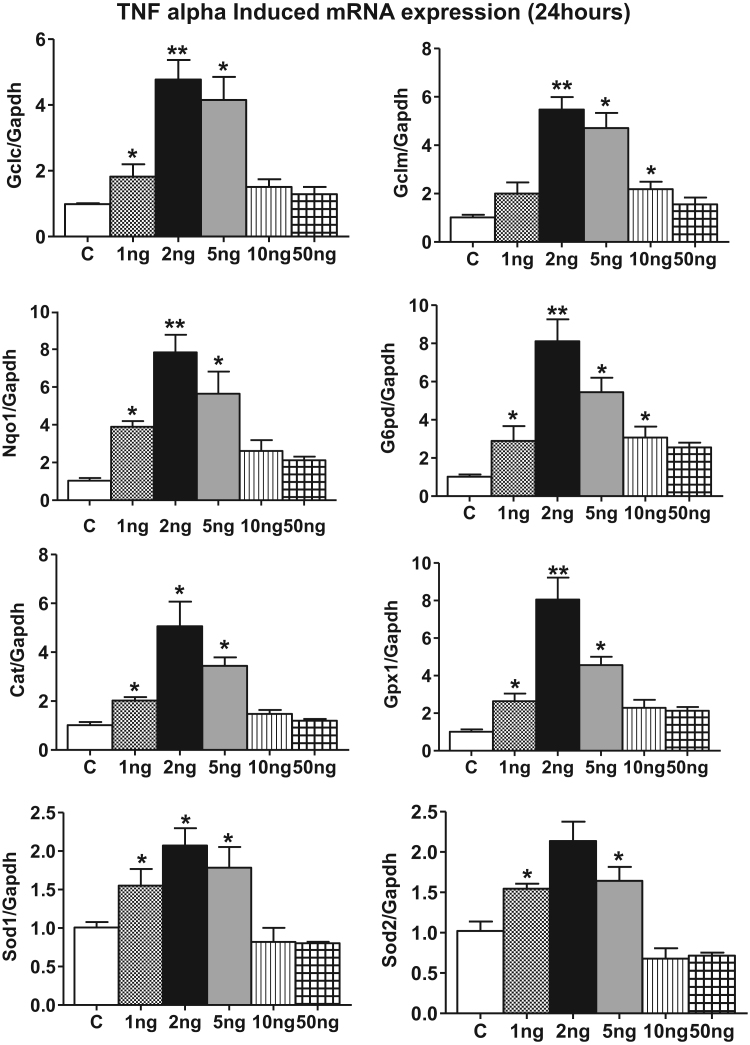
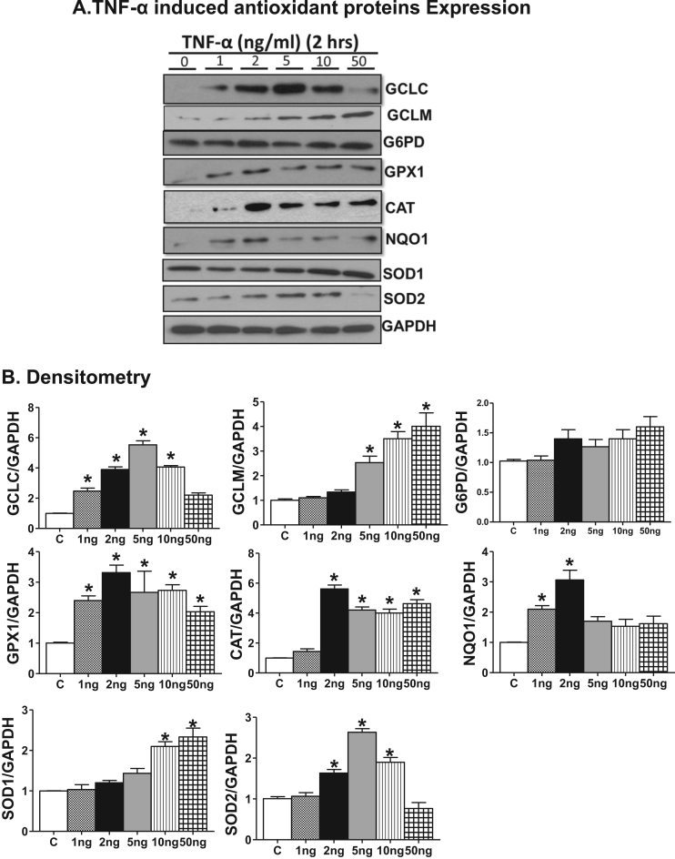
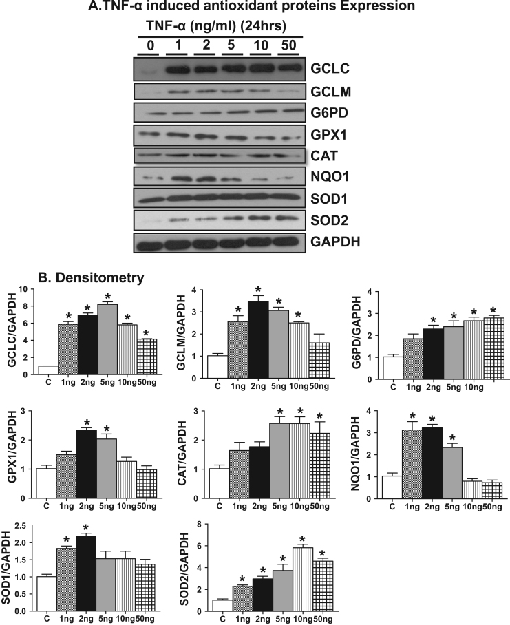
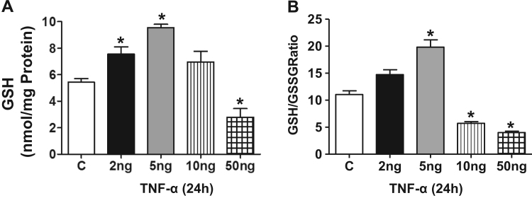
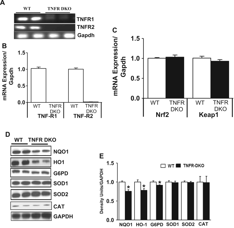
Antagonizing TNF-α signaling attenuates chronic inflammatory disease, but is associated with adverse effects on the cardiovascular system. Therefore the impact of TNF-α on basal control of redox signaling events needs to be understand in more depth. This is particularly important for the Keap1/Nrf2 pathway in the heart and in the present study we hypothesized that inhibition of a low level of TNF-α signaling attenuates the TNF-α dependent activation of this cytoprotective pathway. HL-1 cardiomyocytes and TNF receptor1/2 (TNFR1/2) double knockout mice (DKO) were used as experimental models. TNF-α (2-5ng/ml, for 2h) evoked significant nuclear translocation of Nrf2 with increased DNA/promoter binding and transactivation of Nrf2 targets. Additionally, this was associated with a 1.5 fold increase in intracellular glutathione (GSH). Higher concentrations of TNF-α (>10-50ng/ml) were markedly suppressive of the Keap1/Nrf2 response and associated with cardiomyocyte death marked by an increase in cleavage of caspase-3 and PARP. In vivo experiments with TNFR1/2-DKO demonstrates that the expression of Nrf2-regulated proteins (NQO1, HO-1, G6PD) were significantly downregulated in hearts of the DKO when compared to WT mice indicating a weakened antioxidant system under basal conditions. Overall, these results indicate that TNF-α exposure has a bimodal effect on the Keap1/Nrf2 system and while an intense inflammatory activation suppresses expression of antioxidant proteins a low level appears to be protective.

Keywords: Antioxidants; Apoptosis; Nrf2 signaling; Oxidative stress; TNF-α.


Figures

fx1
Graphical abstract
Fig. 1.
Fig. 1
TNF-α treatment in cell viability and apoptosis in HL-1 cardiomyocytes. HL-1 cells were treated with TNF-α 1, 2, 5, 10, 50 ng/ml for 24 h. (A) At the end of experiment, cells were stained with annexin-V/FITC and read immediately by flow cytometry to measure the extent of apoptosis (n=3). Quantification of percentage live cells at each concentration of TNF-α (B) and percentage dead cells (C) relative to that of control. (D) HL-1 cells treated with TNF-α as in Panel A and protein expression for cleaved fragments of caspase-3 and PARP was determined by immunoblotting. '+' indicates Staurosporine, a positive control. Equal loading was analyzed by anti-GAPDH. Statistical significance was calculated by Mann Whitney test, where *p<0.05 vs untreated control.
Fig. 2.
Fig. 2
TNF-α induced ROS levels in HL-1 cardiomyocytes by H2DCFDA. (A) HL-1 cells were treated with indicated concentrations of TNF-α (as in panel 1 A) and immunofluorescence analysis was performed using H2DCFDA (green fluorescence, 5 µM). The cells were visualized using fluorescence microscopy and images captured using 20× magnification. (B) Three different fields were randomly counted for green positive cells using Image J and the average fluorescence intensity of each concentration of TNF-α relative to that of control was depicted. Statistical significance was calculated by Mann Whitney test, where *p<0.05 vs control. For interpretation of the references to color in this figure legend, the reader is referred to the web version of this article.
Fig. 3.
Fig. 3
TNF-α treatment on Nrf2 Nuclear translocation in HL-1 cardiomyocytes. (A) Representative immunofluorescence photomicrograph (original magnification, 20×) from control and 2 h and 24 h TNFα-treated HL-1 cells (2, 5, 10, 50 ng/ml) showing Nrf2 nuclear localization. DAPI was used as a nuclear counterstain. Data are representative of 3 independent experiments. Relative fluorescence intensity was calculated for Nrf2 nuclear translocation for (B) 2 h and (C) 24 h. Statistical significance was determined by Mann Whitney test, where *p<0.05 compared with untreated control. For interpretation of the references to color in this figure legend, the reader is referred to the web version of this article.
Fig. 4.
Fig. 4
Nrf2 binding activity and mRNA expression in HL-1 cardiomyocytes by TNF-α treatment. Nrf2- DNA binding activity was measured using Trans-AM Nrf2 kit in control and TNF-α treated HL-1 cardiomyocytes (n=3). (D) 2 h and (E) 24 h. Nrf2 gene expression determined in TNF-α treated HL-1 samples for (F) 2 h and (G) 24 h by qPCR. The relative gene expression was calculated by normalizing the mRNA levels of Nrf2 with the levels of GAPDH. In panels, d-G, statistical significance was determined by Mann Whitney test, where *p<0.05 compared with untreated control.
Fig. 5.
Fig. 5
Increased Nrf2 regulated antioxidant genes in HL-1 cardiomyocytes by 24 h of TNF-α treatment. Nrf2 regulated antioxidant Gene expression Profiles (Gclc, Gclm, G6pd, Gpx1, Cat, Nqo1, Sod1 and Sod2) in TNF-α treated HL-1 cell samples for 24 h were analyzed by quantitative PCR and gene levels were normalized with Gapdh (n=3). Mann Whitney test was used to establish statistical significance, where *p<0.05 vs control.
Fig. 6.
Fig. 6
Lower concentration of TNF-α treatment for 2 h increases Nrf2 regulated antioxidant proteins in HL-1 cardiomyocytes. (A) Immunoblot analyses of Nrf2 regulated antioxidant proteins (GCLC, GCLM, G6PD, GPX1, CAT, NQO1, SOD1 and SOD2) in 2 h of TNFα treated HL-1 cells (n=3). (B) Relative intensity of the protein signal was calculated using Image-J software and normalized to GAPDH. Statistical significance was calculated by Mann Whitney test, where *p<0.05 vs control.
Fig. 7.
Fig. 7
TNF-α treatment for 24 h increases Nrf2 regulated antioxidant proteins in HL-1 cardiomyocytes. (A) Immunoblots showing protein expression of antioxidant enzymes in TNF-α treated HL-1 cells (GCLC, GCLM, G6PD, GPX1, CAT, NQO1, SOD1 and SOD2) for 24 h (n=3). (B) Densitometry quantification for appropriate immunoblots normalized with GAPDH intensity. Statistical significance among different treatments were analyzed by Mann Whitney test, where *p<0.05 vs control.
Fig. 8.
Fig. 8
TNF-α induces GSH synthesis in HL-1 cardiomyocytes. (A) HL-1 cells were treated with 2, 5, 10, 50 ng/ml of TNF-α and after 24 h, the cells were processed for quantification of GSH levels (n=3), (B) The GSH/GSSG ratio was calculated from GSH, GSSG values and represented as a bar graph (n=3). Mann Whitney test was performed to establish statistical significance, where *p<0.05 vs control.
Fig. 9.
Fig. 9
TNF-α receptors knockout animals showed decreased basal antioxidant gene/proteins in the heart. (A) One step RT-PCR analysis of TNF-α receptors (TNFR1 and TNFR2) and housekeeping gene Gapdh were determined in WT and TNFR DKO heart samples. (B) Quantitative real time PCR analysis of TNFα receptors (TNFR1 and TNFR2) and housekeeping gene Gapdh were determined in WT and TNFR DKO heart samples (n=4). (C) Nrf2 and Keap1 gene expression profiles were measured by qPCR. Relative gene expression was calculated by normalizing the mRNA levels of gene of interest with Gapdh. (D) Nrf2 regulated antioxidant proteins (NQO1, HO-1, G6PD, SOD1, SOD2 and CAT) were determined by Immunoblot analysis in WT and DKO heart tissue homogenates (n=4). (E) Densitometric analysis of proteins normalized with GAPDH intensity using Image J. Statistical differences were determined by Mann Whitney test, where *p<0.05 vs control.

Similar articles

Cited by

References

    1. Haddad J.J. Antioxidant and prooxidant mechanisms in the regulation of redox(y)-sensitive transcription factors. Cell Signal. 2002;14:879–897. - PubMed
    1. Korashy H.M., El-Kadi A.O. The role of redox-sensitive transcription factors NF-kappaB and AP-1 in the modulation of the Cyp1a1 gene by mercury, lead, and copper. Free Radic. Biol. Med. 2008;44:795–806. - PubMed
    1. Rahman I., Adcock I.M. Oxidative stress and redox regulation of lung inflammation in COPD. Eur. Respir. J. 2006;28:219–242. - PubMed
    1. Maillet A., Pervaiz S. Redox regulation of p53, redox effectors regulated by p53: a subtle balance. Antioxid. Redox Signal. 2012;16:1285–1294. - PubMed
    1. Lee J.M., Li J., Johnson D.A., Stein T.D., Kraft A.D., Calkins M.J., Jakel R.J., Johnson J.A. Nrf2, a multi-organ protector? FASEB J.: Off. Publ. Fed. Am. Soc. Exp. Biol. 2005;19:1061–1066. - PubMed

Publication types

MeSH terms