

Orbital Angiogenesis and Lymphangiogenesis in Thyroid Eye Disease: An Analysis of Vascular Growth Factors with Clinical Correlation
- PMID: 27423310
- PMCID: PMC5536972
- DOI: 10.1016/j.ophtha.2016.05.052
Orbital Angiogenesis and Lymphangiogenesis in Thyroid Eye Disease: An Analysis of Vascular Growth Factors with Clinical Correlation
Abstract
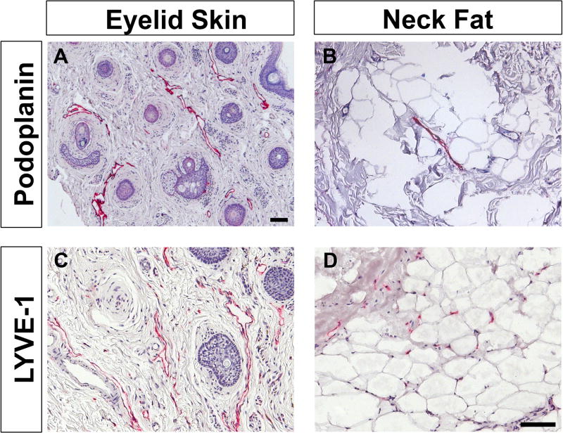
Purpose: The human orbit is an environment that is vulnerable to inflammation and edema in the setting of autoimmune thyroid disease. Our study investigated the tenet that orbital adipose tissue lacks lymphatic vessels and analyzed the clinicopathologic differences between patients with acute and chronic thyroid eye disease (TED). The underlying molecular mediators of blood and lymphatic vessel formation within the orbital fat also were evaluated.
Design: Retrospective cohort study.
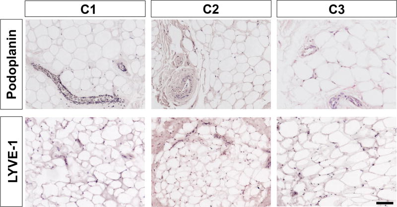
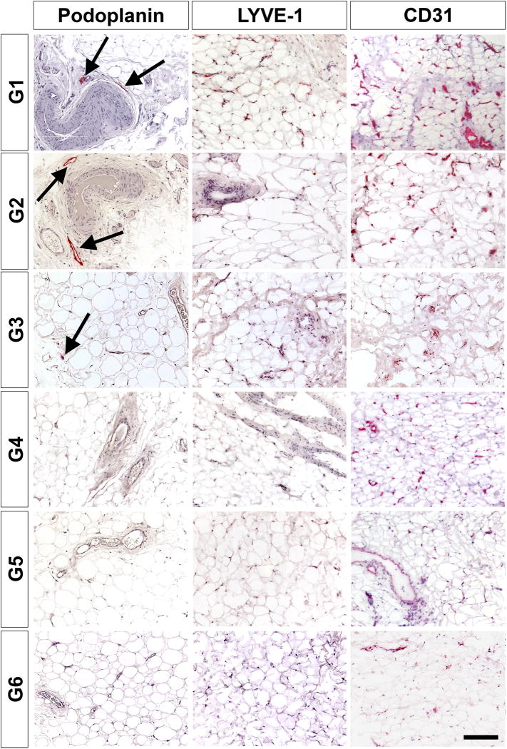
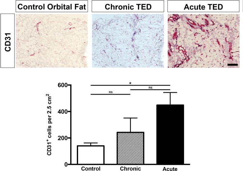
Participants: The study included fat specimens from 26 orbits of 15 patients with TED undergoing orbital decompression. Orbital fat specimens from patients without TED as well as cadaveric orbital fat served as controls.
Methods: Tissue specimens were processed as formalin-fixed, paraffin-embedded sections or frozen cryosections for immunohistochemistry. Total RNA was extracted and analyzed via quantitative (real-time) reverse-transcription polymerase chain reaction. Clinicopathologic correlation was made by determining the clinical activity score (CAS) of each patient with TED.
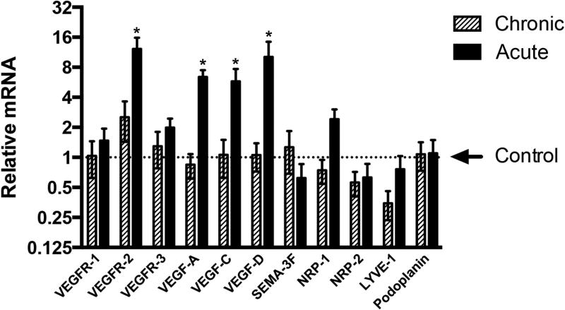
Main outcome measures: Samples were examined for vascular and lymphatic markers including podoplanin, lymphatic vessel endothelial hyaluronan receptor 1 (LYVE-1), and cluster of differentiation 31 (CD31) by immunohistochemistry, as well as for mRNA levels of vascular endothelial growth factor (VEGF), VEGF receptors, semaphorin 3F, neuropilin 1, neuropilin 2, podoplanin, and LYVE-1 by quantitative (real-time) reverse-transcription polymerase chain reaction.
Results: Clinicopathologic correlation revealed increased staining of CD31-positive blood vessels in patients with acute TED with a CAS more than 4, as well as rare staining of podoplanin-positive lymphatic vessels within acutely inflamed orbital fat tissue. Additionally, quantitative (real-time) reverse-transcription polymerase chain reaction analysis demonstrated increased expression of VEGF receptor (VEGFR) 2 as well as VEGF signaling molecules VEGF-A, VEGF-C, and VEGF-D.
Conclusions: In acute TED, compared with chronic TED and control orbital fat, there is increased blood vessel density, suggesting neovascularization and rare lymphatic vessels suggestive of limited lymphangiogenesis. This proangiogenic and prolymphangiogenic microenvironment is likely the result of the increased expression of VEGFR-2, VEGF-A, VEGF-C, and VEGF-D. These findings imply that orbital edema in acute TED may be mediated, in part, by both the formation of new, immature blood vessels and the formation of lymphatic capillaries that are functionally incapable of draining interstitial fluid.
Copyright © 2016 American Academy of Ophthalmology. Published by Elsevier Inc. All rights reserved.
Conflict of interest statement
Figures
Similar articles
-
Increasing lymphatic microvessel density in primary pterygia.Arch Ophthalmol. 2012 Jun;130(6):735-42. doi: 10.1001/archophthalmol.2012.293. Arch Ophthalmol. 2012. PMID: 22801834
-
Vascular endothelial growth factors C and D and their VEGFR-2 and 3 receptors in blood and lymphatic vessels in healthy and arthritic synovium.J Rheumatol. 2002 Jan;29(1):39-45. J Rheumatol. 2002. PMID: 11824969
-
Intracranial meningiomas, the VEGF-A pathway, and peritumoral brain oedema.Dan Med J. 2013 Apr;60(4):B4626. Dan Med J. 2013. PMID: 23651727 Review.
-
The angiogenic switch for vascular endothelial growth factor (VEGF)-A, VEGF-B, VEGF-C, and VEGF-D in the adenoma-carcinoma sequence during colorectal cancer progression.J Pathol. 2003 Jun;200(2):183-94. doi: 10.1002/path.1339. J Pathol. 2003. PMID: 12754739
-
Lymphangiogenic growth factors, receptors and therapies.Thromb Haemost. 2003 Aug;90(2):167-84. doi: 10.1160/TH03-04-0200. Thromb Haemost. 2003. PMID: 12888864 Review.
Cited by
-
Future Projections in Thyroid Eye Disease.J Clin Endocrinol Metab. 2022 Aug 8;107(Suppl_1):S47-S56. doi: 10.1210/clinem/dgac252. J Clin Endocrinol Metab. 2022. PMID: 36346684 Free PMC article. Review.
-
A novel therapy for fracture healing by increasing lymphatic drainage.J Orthop Translat. 2024 Mar 13;45:66-74. doi: 10.1016/j.jot.2024.02.001. eCollection 2024 Mar. J Orthop Translat. 2024. PMID: 38511124 Free PMC article.
-
miR-199a Downregulation as a Driver of the NOX4/HIF-1α/VEGF-A Pathway in Thyroid and Orbital Adipose Tissues from Graves' Patients.Int J Mol Sci. 2021 Dec 23;23(1):153. doi: 10.3390/ijms23010153. Int J Mol Sci. 2021. PMID: 35008579 Free PMC article.
-
Transient Expression of Lymphatic Markers in Retrobulbar Intraconal Orbital Vasculature During Fetal Development.Invest Ophthalmol Vis Sci. 2020 Jun 3;61(6):22. doi: 10.1167/iovs.61.6.22. Invest Ophthalmol Vis Sci. 2020. PMID: 32516408 Free PMC article.
-
Effect of Methotrexate on an In Vitro Patient-Derived Model of Proliferative Vitreoretinopathy.Invest Ophthalmol Vis Sci. 2017 Aug 1;58(10):3940-3949. doi: 10.1167/iovs.16-20912. Invest Ophthalmol Vis Sci. 2017. PMID: 28777835 Free PMC article.
References
Publication types
MeSH terms
Substances
Grants and funding
LinkOut - more resources
Full Text Sources
Other Literature Sources
Medical
Miscellaneous
