. 2016 Jul 31;428(15):3131-46.
doi: 10.1016/j.jmb.2016.07.003. Epub 2016 Jul 14.

Chopping and Changing: the Evolution of the Flavin-dependent Monooxygenases

Affiliations

Chopping and Changing: the Evolution of the Flavin-dependent Monooxygenases

Maria Laura Mascotti et al. J Mol Biol. .

Abstract

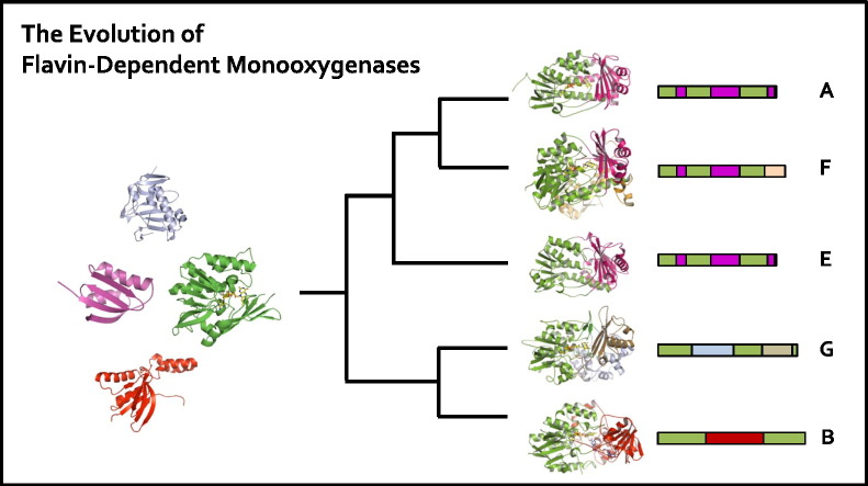
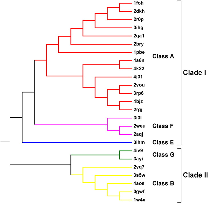
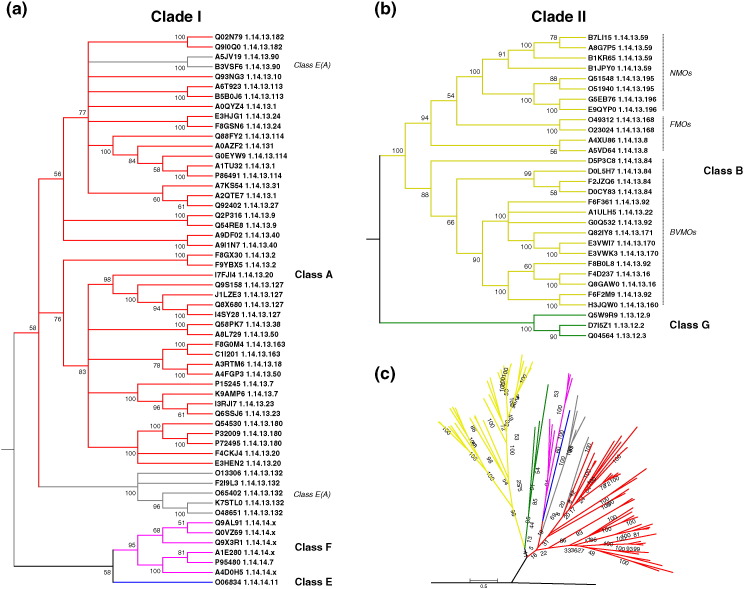
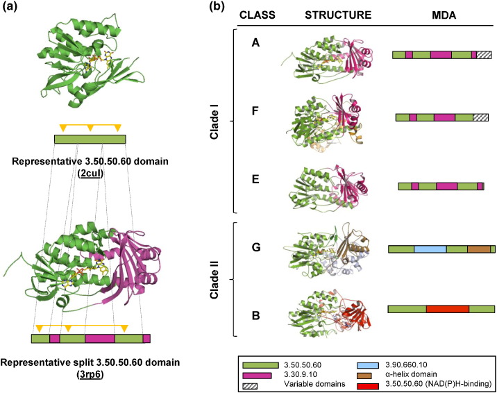
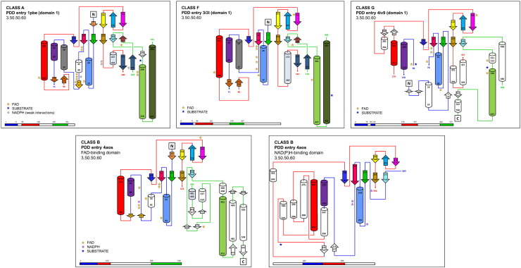
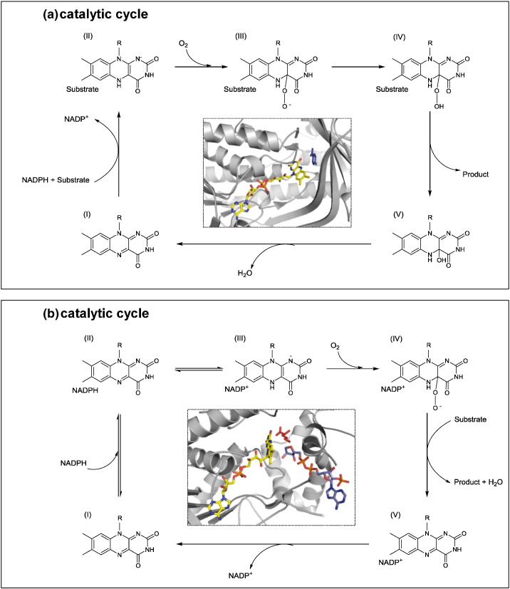
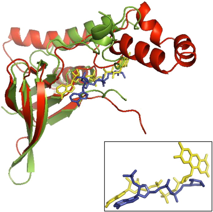
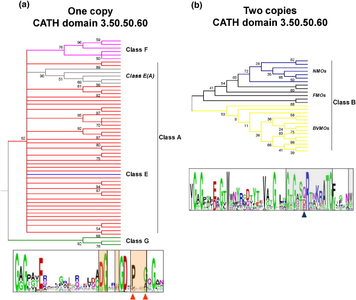
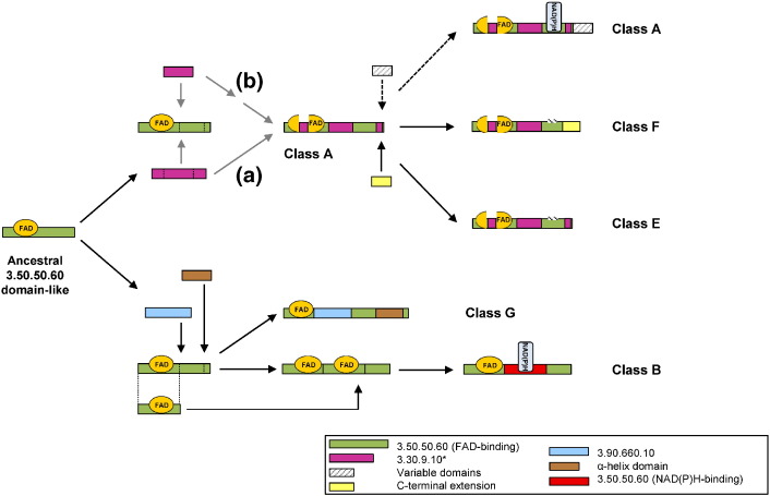
Flavin-dependent monooxygenases play a variety of key physiological roles and are also very powerful biotechnological tools. These enzymes have been classified into eight different classes (A-H) based on their sequences and biochemical features. By combining structural and sequence analysis, and phylogenetic inference, we have explored the evolutionary history of classes A, B, E, F, and G and demonstrate that their multidomain architectures reflect their phylogenetic relationships, suggesting that the main evolutionary steps in their divergence are likely to have arisen from the recruitment of different domains. Additionally, the functional divergence within in each class appears to have been the result of other mechanisms such as a complex set of single-point mutations. Our results reinforce the idea that a main constraint on the evolution of cofactor-dependent enzymes is the functional binding of the cofactor. Additionally, a remarkable feature of this family is that the sequence of the key flavin adenine dinucleotide-binding domain is split into at least two parts in all classes studied here. We propose a complex set of evolutionary events that gave rise to the origin of the different classes within this family.

Keywords: cofactor-binding enzymes; enzyme evolution; flavin-dependent monooxygenases; multidomain architecture.


Figures

Image 1
Graphical abstract
Fig. 1
Fig. 1
Structure-based phylogeny of the Group 1 flavin-dependent monooxygenases. Molecular phylogenetic analysis by the ML method based on the 3D alignment of the whole 29 available Group 1 protein structures. The PDB codes of each structure are given on the right. The BS consensus tree inferred from 100 replicates was taken to represent the evolutionary history of the taxa analyzed. Branches corresponding to partitions reproduced in fewer than 50% BS replicates are collapsed. The same clade composition and tree topology were obtained when using Bayesian inference. Classes of flavin-dependent monooxygenases are shown in different colors as follows: class A (red), class F (hot pink), class E (blue), class G (green), and class B (yellow).
Fig. 2
Fig. 2
Sequence-based phylogeny of the Group 1 flavin-dependent monooxygenases. Molecular phylogenetic analysis by the ML method from MSAs of manually edited amino acid subsequences corresponding to the FAD-binding domain (CATH 3.50.50.60) only. BS values are indicated at the branches. Branches corresponding to partitions reproduced in fewer than 50% BS replicates are collapsed. UniProt codes and EC numbers are given for each sequence. (a) The phylogenetic tree of the classes included in the first clade of the structure-based tree (Fig. 1). (b) The evolutionary relationships for the second clade in Fig. 1. (c) The phylogenetic tree of all classes of Group 1 flavin‐monooxygenases. The sequence of precorrin synthase (UniProt code: D5AUZ5) was used as an external group to root the tree. Classes of flavin-dependent monooxygenases are shown in different colors: class A (red), class E(A) (grey), class E (blue), class F (hot pink), class B (ochre), and class G (green).
Fig. 3
Fig. 3
An overview of the MDAs in Group 1 flavin-dependent monooxygenases. (a) Structure and schematic MDA bar diagrams for: Top, a single-domain enzyme consisting of a complete CATH 3.50.50.60 domain (PDB entry: 2CUL); and Bottom, a flavin monooxygenase (3RP6) containing a split 3.50.50.60 domain (green) merged with a split domain 3.30.9.10 (purple). The dashes indicate where the boundaries of the split domain occur in the full domain. The FAD contact sites are represented by the yellow triangles. (b) Overview of the MDAs of each class of flavin-dependent enzymes. Colors in the structure correspond to those in the MDA diagrams. The key at the bottom shows the color code for each CATH domain. Note that the green and purple domains are each a single domain that has been split into segments of noncontiguous amino acids.
Fig. 4
Fig. 4
Topology diagrams of the FAD-binding domain. Topology diagrams of the 3.50.50.60 CATH domain in each of the Group 1 classes. Helices are depicted as cylinders and β-strands as arrows. The colors represent secondary structure elements that overlap when the 3D structures are superposed. White elements indicate unique features to the given structure. The stars identify residues involved in the interaction with FAD, NAPH, and substrate. The location of each domain in the protein's MDA is highlighted in the schematic bar diagrams at the bottom. The colors of the bars correspond to the colors of the connecting lines joining the secondary structure elements in the corresponding topology diagram.
Fig. 5
Fig. 5
Catalytic mechanisms of class A and class B monooxygenases. Top: schematic representation of the catalytic cycle of class A monooxygenases. The cycle initiates when the ES complex is formed (I) and the oxidized flavin is reduced by NADPH, then the reaction with molecular oxygen takes place to form the C4a-peroxyflavin and C4a-flavin hydroperoxide species (III and IV, respectively); finally, the substrate is oxidized and the product released. At the center, the structure of the active site of PHBH enzyme (PDB entry: 1PBE) is shown, FAD is presented as yellow sticks and substrate p-hydroxybenzoate in dark blue. Bottom: as above but for class B monooygenases. Catalytic cycle starts with the recruitment of NADPH (I → II) and the subsequent flavin reduction (III). Reduced enzyme reacts with molecular oxygen to form the key intermediate C4-a peroxyflavin (IV), which then transforms the substrate into the product. NADP+ remains bound during the whole catalytic cycle and the rate-determining step in the releasing of this oxidized cofactor (V → I). In the center, the structure of active site of enzyme ONMO is shown (PDB entry: 3S5W), FAD is presented as yellow sticks, NADPH in light blue, and substrate L-ornithine in red.
Fig. 6
Fig. 6
Class B nucleotide-binding domains. Superposition of the first half of a split 3.50.50.60 CATH domain (green) involved in binding FAD (yellow sticks) and an unsplit 3.50.50.60 CATH domain (red) involved in NADPH binding (light blue sticks). Both domains come from PDB entry 3S5W. The RMSD between equivalent Cα atoms is 2.26 Å. The inset at bottom right shows just the cofactors FAD and NADPH from the superposition, demonstrating that they both bind in the same equivalent position and orientation in their respective domain.
Fig. 7
Fig. 7
The origin of enzyme function in flavin-dependent monooxygenases. (a) Top: phylogenetic tree of flavin monooxygenases containing a single CATH 3.50.50.60 domain (FAD-binding domain). The tree was constructed from an MSA of subsequences corresponding to just the 39 residues involved in interactions with cofactors and substrates. The ML inference method was used, and BS values are indicated at the branches. Branches corresponding to fewer than 50% BS replicates are collapsed. Colored branches show the different flavin monooxygenases classes: class A (red), class E(A) (grey), class E (blue), class F (hot pink), and class G (green). Bottom: logo scheme employed for the MSA; residues in contact with FAD (empty boxes), with substrate (light orange boxes), and residues in contact with both FAD and the substrate are indicated by orange triangles. (b) Top: as above but for class B enzymes that contain two CATH 3.50.50.60 domains (FAD- and NADPH-binding domains). Here, the colored branches indicate the different subtypes within class B: BVMOs (ochre), NMOs (dark blue), and FMOs (black). Bottom: logo scheme employed for the MSA; residues in contact with FAD (empty boxes) andNAD(P)H (grey boxes), while residues in contact with FAD and NAD(P)H are identified by a blue triangle.
Fig. 8
Fig. 8
Graphical representation of the proposed evolution and divergence of flavin-dependent monooxygenases. The figure shows the proposed evolutionary events at the MDA level that led from a common FAD-binding ancestor to the emergence of the five classes A, B, E, F, and G of the flavin monooxygenases. Two main divergence events are represented: Top, the fusion to the 3.30.9.10 CATH domain, and the concomitant split of the 3.50.50.60 CATH domain originated classes A, F, and E, alternative (a). The asterisked domain (CATH 3.30.9.10), which, according to Gene3D, is a split domain, is more likely to consist of a single 3.30.70.100 domain at the center and two smaller embellishments, corresponding to the outer parts (see main text). This alternative scenario is presented as (b). Class A is proposed to acquire the ability of NADPH binding after a complex set of structural changes. This class may present an extra C-terminal variable domain (indicated by the dashed arrows). Class F emerged after the recruitment of a C-terminal terminal extension, while class E seems to remain similar to the original common structure. These last two classes lack the ability of binding NADPH. Bottom, class G monooxygenases originated after the recruitment of a CATH domain 3.90.660.10 (involved in substrate binding) and a C-terminal α-helix with the concomitant split of the CATH domain 3.50.50.60 into three parts. On the other hand, class B emerged after a sequence of events involving the partial duplication of the FAD-binding domain and the insertion of this duplicated domain into the original one followed by a change in its specificity toward the binding of NADPH. The inset shows the color code for each CATH domain.

References

    1. Macheroux P., Kappes B., Ealick S.E. Flavogenomics—a genomic and structural view of flavin-dependent proteins. FEBS J. 2011;278:2625–2634. - PubMed
    1. Mattevi A. To be or not to be an oxidase: challenging the oxygen reactivity of flavoenzymes. Trends Biochem. Sci. 2006;31:276–283. - PubMed
    1. Balke K., Kadow M., Mallin H., Sass S., Bornscheuer U.T. Discovery, application and protein engineering of Baeyer–Villiger monooxygenases for organic synthesis. Org. Biomol. Chem. 2012;10:6249–6265. - PubMed
    1. Mascotti M.L., Juri Ayub M., Dudek H., Sanz M.K., Fraaije M.W. Cloning, overexpression and biocatalytic exploration of a novel Baeyer–Villiger monooxygenase from Aspergillus fumigatus Af293. AMB Express. 2013;3:33. - PMC - PubMed
    1. Riebel A., Dudek H.M., de Gonzalo G., Stepniak P., Rychlewski L., Fraaije M.W. Expanding the set of rhodococcal Baeyer–Villiger monooxygenases by high-throughput cloning, expression and substrate screening. Appl. Microbiol. Biotechnol. 2012;95:1479–1489. - PMC - PubMed

Publication types

LinkOut - more resources